. 2011 Apr;49(4):278-94.
doi: 10.1002/dvg.20707. Epub 2011 Mar 5.

Molecular analysis of neurogenic placode development in a basal ray-finned fish

Affiliations

Molecular analysis of neurogenic placode development in a basal ray-finned fish

Melinda S Modrell et al. Genesis. 2011 Apr.

Abstract

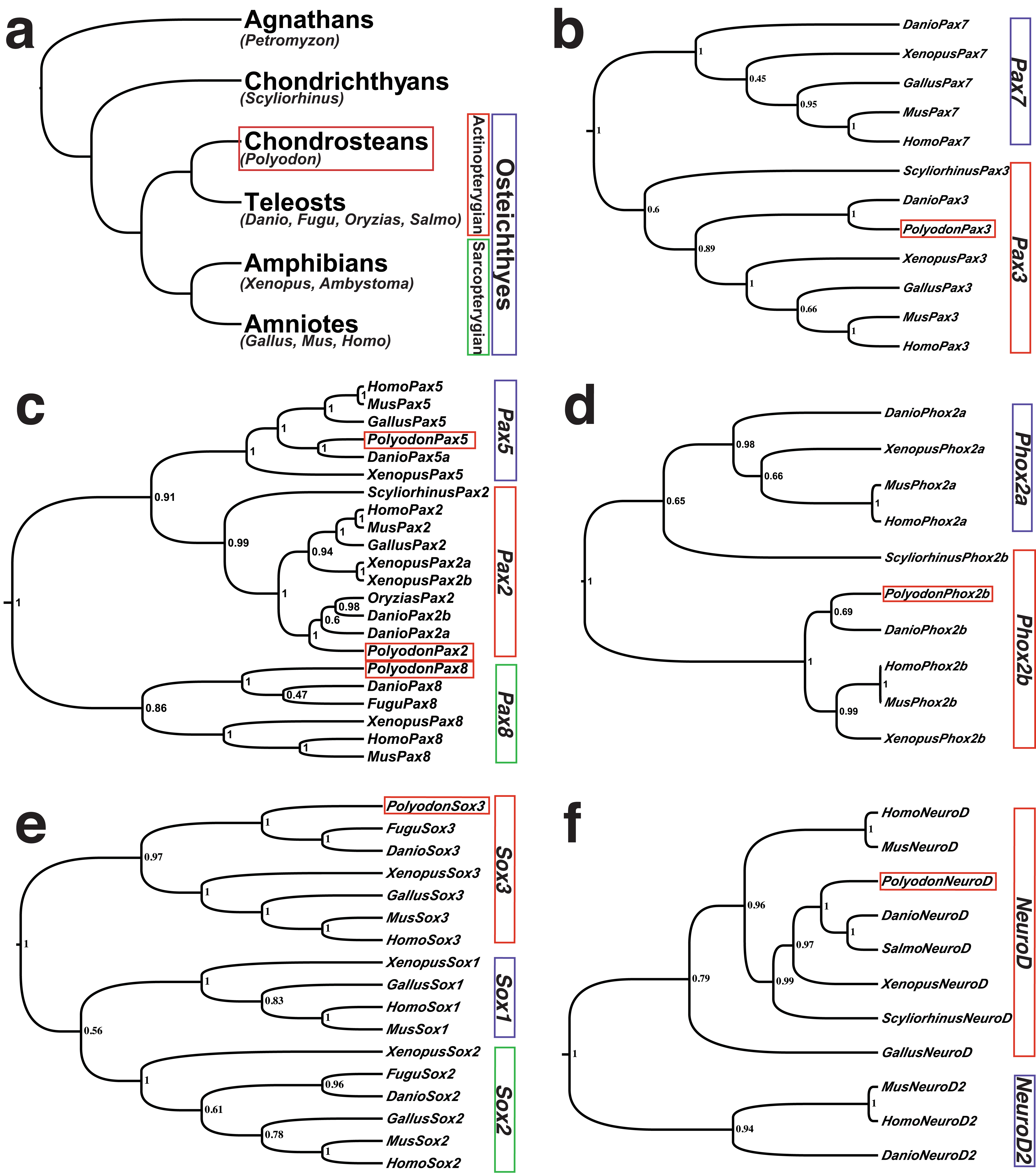
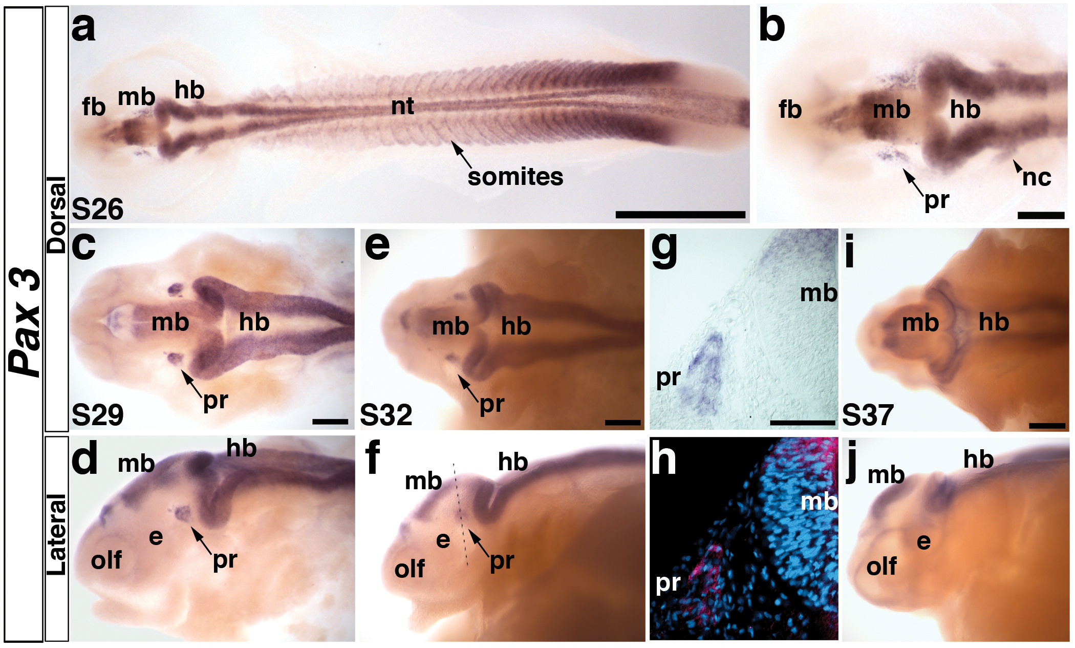
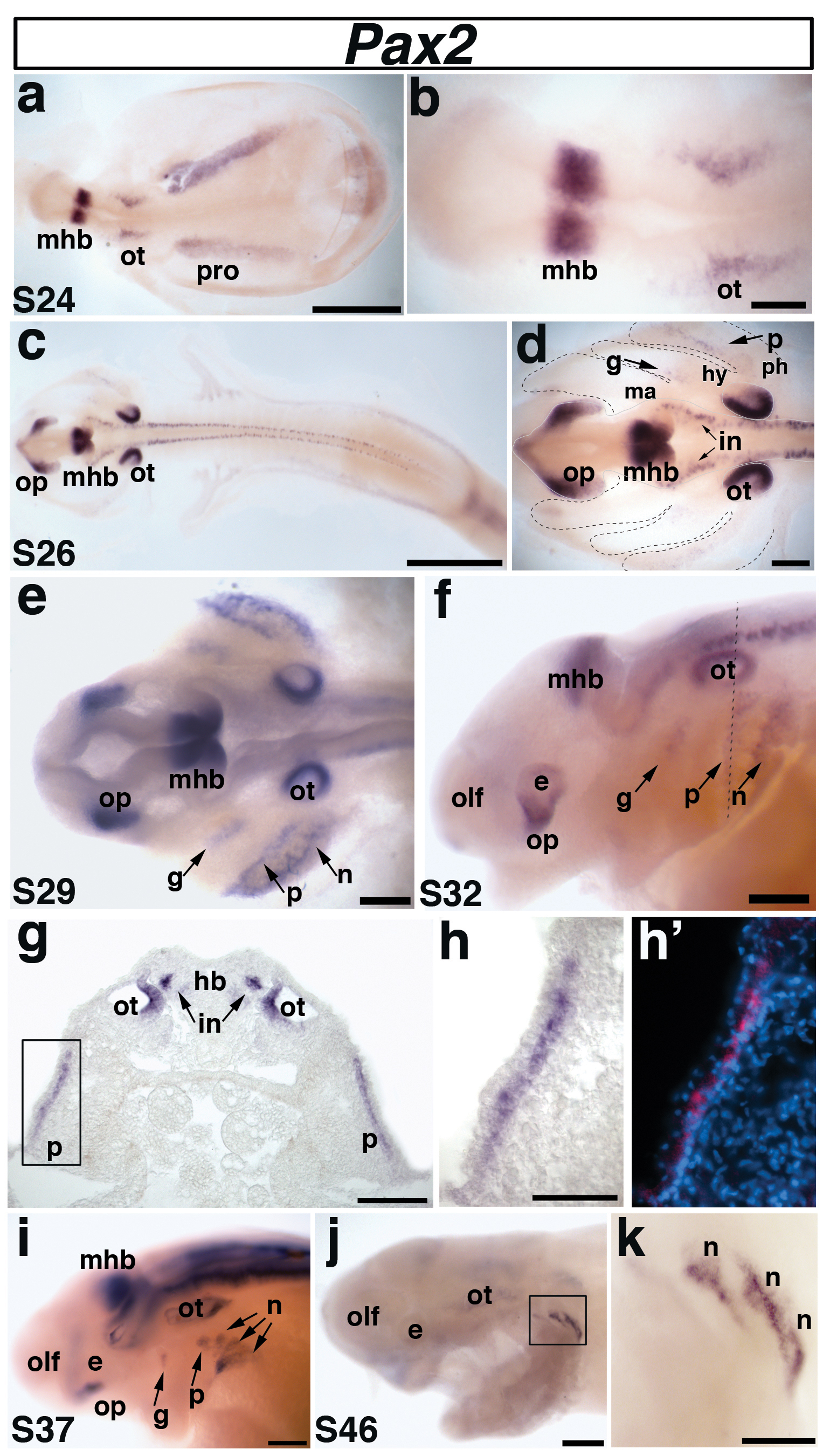
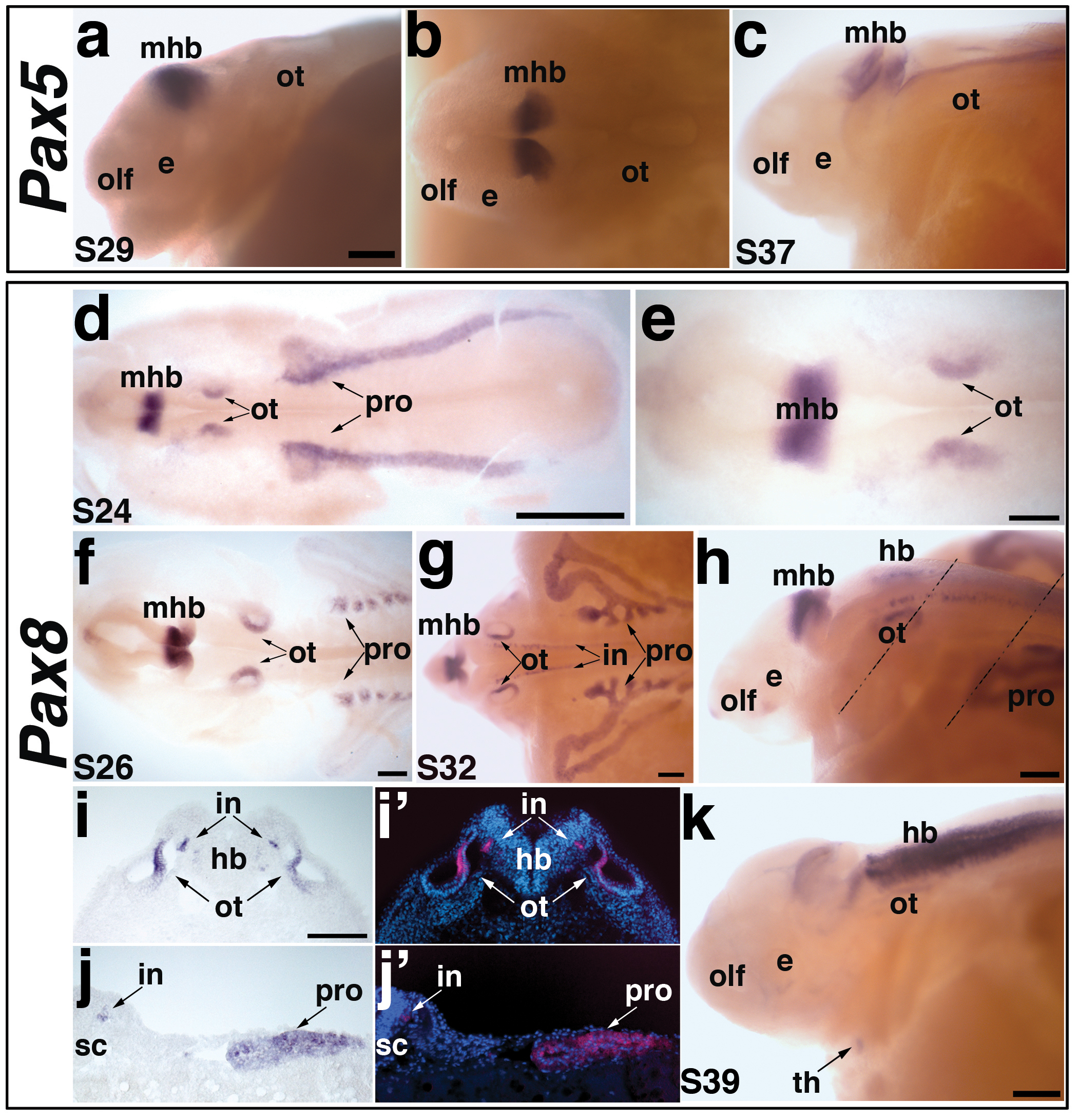
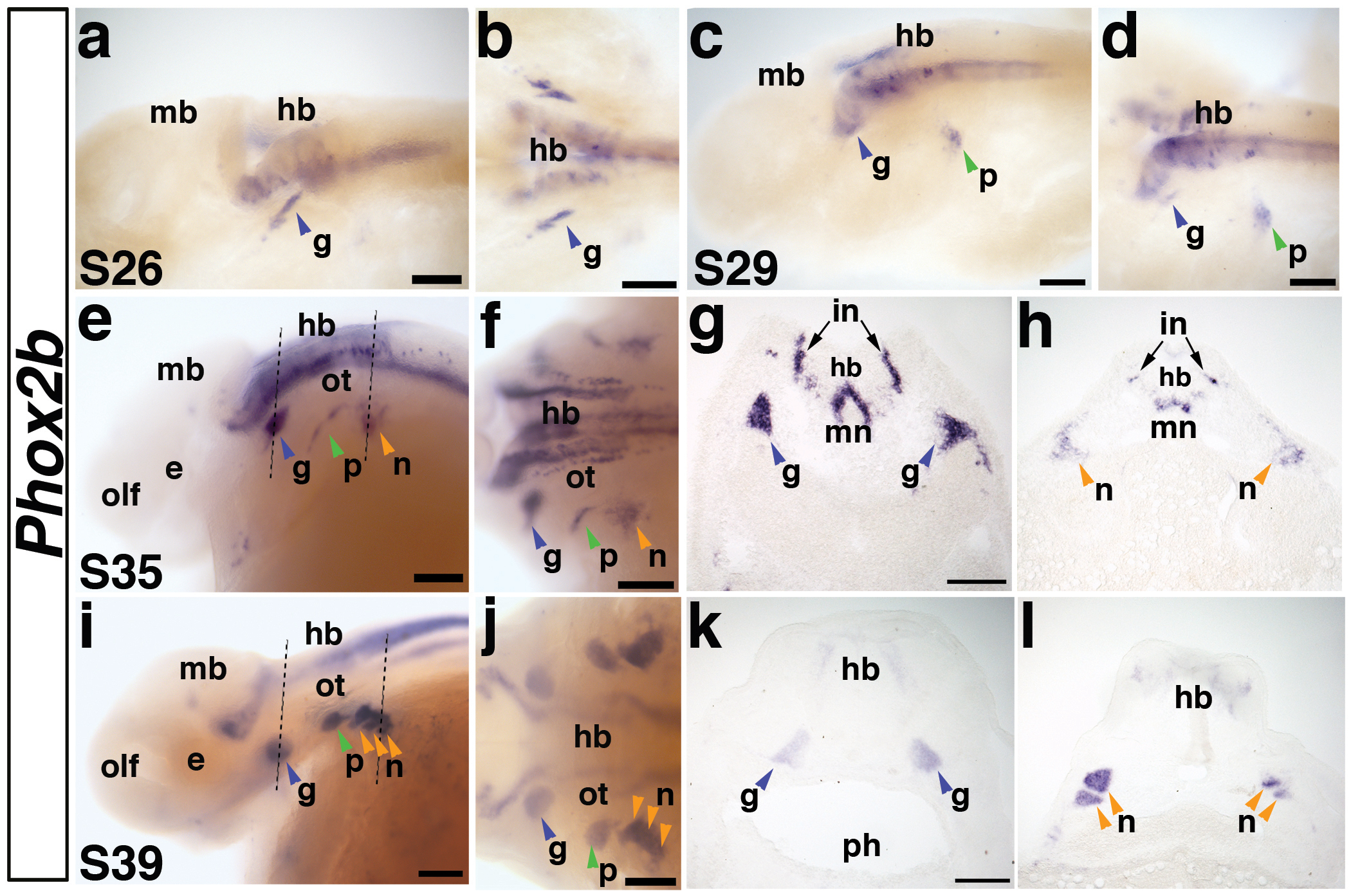
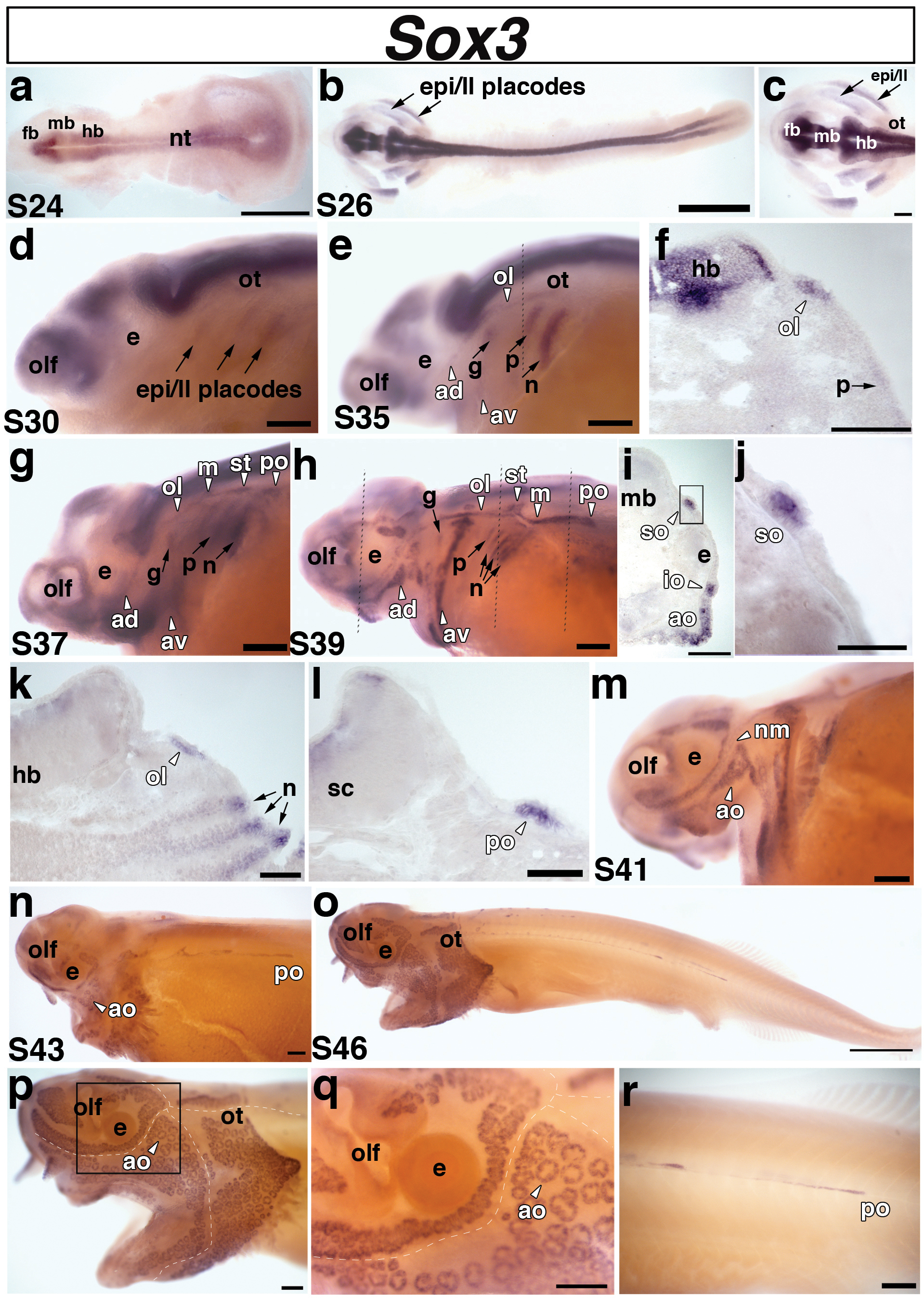
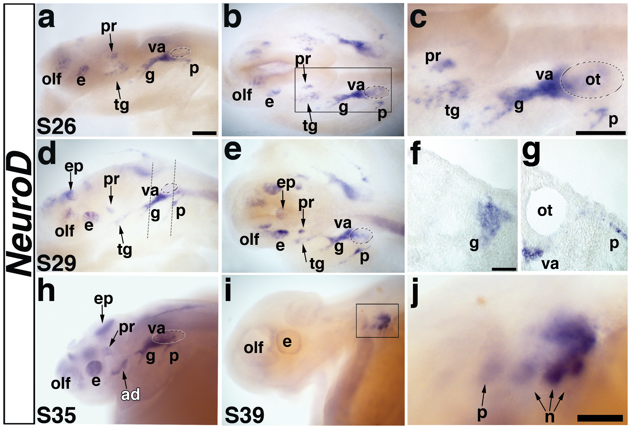
Neurogenic placodes are transient, thickened patches of embryonic vertebrate head ectoderm that give rise to the paired peripheral sense organs and most neurons in cranial sensory ganglia. We present the first analysis of gene expression during neurogenic placode development in a basal actinopterygian (ray-finned fish), the North American paddlefish (Polyodon spathula). Pax3 expression in the profundal placode confirms its homology with the ophthalmic trigeminal placode of amniotes. We report the conservation of expression of Pax2 and Pax8 in the otic and/or epibranchial placodes, Phox2b in epibranchial placode-derived neurons, Sox3 during epibranchial and lateral line placode development, and NeuroD in developing cranial sensory ganglia. We identify Sox3 as a novel marker for developing fields of electrosensory ampullary organs and for ampullary organs themselves. Sox3 is also the first molecular marker for actinopterygian ampullary organs. This is consistent with, though does not prove, a lateral line placode origin for actinopterygian ampullary organs.


Figures

Fig. 1
Fig. 1. Phylogenetic trees generated using a Bayesian-coalescence framework confirm the orthology of cloned Polyodon spathula (paddlefish) cDNA fragments
(a) A highly simplified vertebrate phylogeny showing the relationship of the main vertebrate developmental and genetic model systems in relation to the chondrostean (basal acintopterygian) Polyodon spathula (North American paddlefish) (red box), as well as other basal vertebrates used in comparative developmental studies, such as the lamprey Petromyzon marinus (an agnathan, i.e., jawless fish), and the shark Scyliorhinus canicula (a chondrichthyan, i.e., cartilaginous fish). Tetrapods (amphibians and amniotes) and teleosts are members, respectively, of the two sister groups of bony fish: sarcopterygians (lobe-finned fish) and actinopterygians (ray-finned fish). (b-f) Phylogenetic trees showing the orthology of the Polyodon gene fragment with other vertebrates. Polyodon sequences are outlined by a red box. Numbers at nodes represent the Bayesian posterior probability of each clade. (b) Pax3. (c) Pax2/5/8 family. (d) Phox2b. (e) Sox3. (f) NeuroD. Species abbreviations are as follows: Danio, Danio rerio (zebrafish); Fugu, Takifugu rubripes (pufferfish); Gallus, Gallus gallus (chicken); Homo, Homo sapiens (human); Mus, Mus musculus (mouse); Oryzias, Oryzias latipes (medaka); Polyodon, Polyodon spathula (paddlefish); Salmo, Salmo salmo (salmon); Scyliorhinus, Scyliorhinus canicula (lesser-spotted dogfish/catshark); Xenopus, Xenopus laevis (African clawed frog).
Fig. 2
Fig. 2. Paddlefish Pax3 is expressed in the profundal placode and ganglion
(a) Embryo at stage 26 showing the distribution of Pax3 transcripts in the neural tube, somites, and head. At this stage, the primary divisions of the head into forebrain (fb), midbrain (mb) and hindbrain (hb) are evident. (b) Higher power view of head region in panel a. Pax3 is expressed in bilateral patches of cells lateral to the midbrain (mb) in the profundal placode (pr) and in neural crest cells (nc) migrating from the hindbrain (hb) towards the pharyngeal arches. (c,d) Stage 29 embryo in dorsal and lateral views, respectively. The Pax3-positive cells in the profundal placode (pr) have condensed, yet remain near the epithelium. Pax3 is also weakly expressed in the olfactory pits (olf). (e,f) Stage 32 embryo in dorsal and lateral views. The Pax3-positive cells have delaminated from the epithelium into the mesenchyme in the developing profundal ganglion (pr). Dotted line shows plane of section shown in panels g and h. (g) Brightfield image of a transverse section through the stage 32 embryo shown in panel f. Pax3-positive cells are clearly expressed in the developing profundal ganglion (pr). (h) Corresponding false color overlay image of brightfield image in panel g (red, Pax3) with the nuclear marker DAPI (blue). (i,j) Stage 37 embryo in dorsal and lateral views. Pax3 expression persists in the central nervous system, yet is completely absent in the profundal ganglion by this stage. Abbreviations: e, eye; fb, forebrain hb, hindbrain; mb, midbrain; nt, neural tube; olf, olfactory pits; pr, profundal placode or ganglion; s, stage. Scale bars: (a) 1 mm; (b-f, i, j) 200 μm; (g,h) 50 μm.
Fig. 3
Fig. 3. Paddlefish Pax2 is expressed in the otic and epibranchial placodes
(a) Dorsal view of a stage 24 embryo. Pax2 is expressed in the midbrain-hindbrain boundary (mhb), the otic placode (ot), and the pronephros (pro). (b) Higher power view of the head region in panel a shows Pax2 expression in the otic placode (ot) and midbrain-hindbrain boundary (mhb). (c) Dorsal view of a stage 26 embryo. (d) Higher power view of head region in panel c. Pax2 continues to be strongly expressed in the midbrain-hindbrain boundary (mhb) and the developing otic vesicle (ot), but has been downregulated in the pronephros (pro). New domains of Pax2 expression are also present in the optic stalk (op), and interneurons (in) of the hindbrain and spinal cord, as well as in the developing geniculate (g) and petrosal (p) epibranchial placodes. Pax2 expression highlights the geniculate placode (g), which is forming dorsocaudal to the external position of the first pharyngeal pouch (i.e., future position of the first pharyngeal cleft) between the mandibular (ma) and hyoid (hy) arches (boundaries indicated with black dashed lines). The developing petrosal placode (p) is located dorsocaudal to the future second pharyngeal cleft between the hyoid (hy) and post-hyoid arches (ph). (e) Dorsal view of a stage 29 embryo. Pax2 continues to be expressed in the previously described domains, with stronger expression within the developing epibranchial placodes, now including the nodose placodes (n) developing caudal to the future third pharyngeal cleft. (f). Lateral view of a stage 32 embryo. Pax2 expression continues in all domains. Dotted line shows plane of section shown in panels g-h’. (g-h’) Transverse section through the stage 32 embryo shown in panel f. (g) Pax2 is expressed in the medial region of the otic vesicle (ot), interneurons (in) of the hindbrain (hb), and in the petrosal placode (p). (h) Higher power view of the petrosal placode (p) from panel g in brightfield and (h’) corresponding false color overlay (red) with DAPI (blue). Pax2 is only seen in the deep layer of the stratified epithelial ectoderm, adjacent to the basement membrane. (i) Lateral view of a stage 37 embryo. The expression of Pax2 in the nodose placodes has resolved further into three distinct domains representing individual nodose placodes. (j) Lateral view of a stage 46 embryo and (k) higher power view of boxed area in panel j. Pax2 expression is restricted to the caudalmost nodose placodes. Abbreviations: e, eye; g, geniculate; hb, hindbrain; hy, hyoid arch; in, interneurons; ma, mandibular arch; mhb, midbrain-hindbrain boundary; n, nodose; olf, olfactory pits; op, optic stalk; ot, otic; p, petrosal; ph, post-hyoid arches; pro, pronephros; s, stage. Scale bars: (a,c) 1 mm; (b-f, i,j) 200 μm; (g,k) 100 μm; (h,h’) 50 μm.
Fig. 4
Fig. 4. Paddlefish Pax5 and Pax8 expression during otic placode and pronephros development
(a-c) Pax5 expression in paddlefish. (a,b) A stage 29 embryo, in lateral and dorsal views, respectively, showing Pax5 expression. Pax5 is not expressed in any placodes, but is expressed in the midbrain-hindbrain boundary (mhb). (c) Lateral view of a stage 37 embryo. Pax5 expression is maintained in the midbrain-hindbrain boundary, and more weakly seen in the hindbrain and spinal cord. (d-k) Pax8 expression in paddlefish. (d) Dorsal view of a stage 24 embryo showing Pax8 expression. Pax8 is restricted to the midbrain-hindbrain boundary (mhb), the otic vesicle (ot), and the developing pronephros (pro). (e) Higher power view of head region from panel d, showing Pax8 in the midbrain-hindbrain boundary (mhb) and otic vesicle (ot). (f) Dorsal view of a stage 26 embryo. Pax8 is maintained in the previously described domains. In the elaborating pronephros, Pax8 appears restricted specifically to the pronephric canals, adjacent to the somites. (g,h) Dorsal and lateral views of a stage 32 embryo. Pax8 expression is maintained in the midbrain-hindbrain boundary (mhb), otic vesicle (ot) and pronephros (pro). Pax8 is also strongly expressed in the broadening pronephric canals and ducts and in the interneurons (in) of the hindbrain (hb) and spinal cord (sc). Dotted lines in panel h show planes of sections shown in panels i-i’ and j-j’. (i) Brightfield image and (i’) corresponding false color overlay image (red, Pax8) with DAPI (blue) showing a section through the otic vesicle of the stage 32 embryo shown in panel h. Pax8 is expressed in a medial region of the otic vesicle (ot) and interneurons (in) of the hindbrain (hb). (j) Brightfield image and (j’) corresponding false color overlay image (red, Pax8) with DAPI (blue) showing a section through the pronephros of the stage 32 embryo shown in panel h. Pax8 is observed in the interneurons (in) and pronephric duct (pro). (k). Lateral view of a stage 39 embryo. Pax8 is weakly expressed in the otic vesicle (ot); expression in the hindbrain (hb) has broadened extensively. Pax8 is also expressed in the putative developing thyroid gland (arrow). Abbreviations: e, eye; hb, hindbrain; in, interneurons; mhb, midbrain-hindbrain boundary; olf, olfactory pits; ot, otic; pro, pronephros; s, stage; sc, spinal cord, th, thyroid gland. Scale bars: (a-c) 200 μm; (d) 1 mm; (e-h,k) 200 μm; (i,j) 100 μm.
Fig. 5
Fig. 5. Paddlefish Phox2b is expressed in epibranchial placode-derived neurons
(a,b) Lateral and dorsal views, respectively, of a stage 26 embryo. Phox2b is expressed in a stripe of cells representing the first neurons of the developing geniculate ganglion (g) (blue arrowhead) and in neurons in the developing hindbrain (hb). Expression of Phox2b in the hindbrain (hb) persists throughout embryogenesis. (c,d) Lateral and dorsal views of a stage 29 embryo. Phox2b is expressed in the condensing geniculate ganglion (blue arrowhead) and now also in the developing petrosal ganglion (p) (green arrowhead). (e,f) Lateral and dorsal views of a stage 35 embryo. Phox2b expression continues in the geniculate ganglion (blue arrowhead), petrosal ganglion (green arrowhead), and has begun in a patch of cells representing the developing nodose ganglia (n) (orange arrowhead). Dotted lines in panel e indicate planes of sections shown in panels g and h. (g) Section through the geniculate ganglia (blue arrowheads) of the stage 35 embryo shown in panels e,f. Phox2b transcripts are strongly expressed in the geniculate ganglia, as well as in a subset of hindbrain neurons, presumably including interneurons (in) and motor neurons (mn). (h) Section through the developing nodose ganglia (orange arrow) in the stage 35 embryo shown in panel e. Compared to the condensed geniculate ganglion, Phox2b-positive cells of the developing nodose ganglia are still scattered, such that individual ganglia cannot be distinguished. (i,j) Lateral and dorsal views of a stage 39 embryo. Phox2b expression clearly marks all the condensed epibranchial placode-derived ganglia (arrowheads). Dotted lines in panel i indicate planes of sections in panels k and l. (k) Section through the geniculate ganglia (blue arrowheads) of the stage 39 embryo shown in panels i,j, showing Phox2b expression in the medially located ganglia. (l) Section through the nodose ganglia (orange arrowheads) of the stage 39 embryo shown in panels i,j. At this stage, individual Phox2b-positive nodose ganglia can be distinguished. Abbreviations: e, eye; g, geniculate ganglion; hb, hindbrain; in, interneurons; mb, midbrain; mn, motor neurons; n, nodose ganglia, olf, olfactory pits; ot, otic; p, petrosal ganglion; ph, pharynx; s, stage. Scale bars: (a-f; i-j) 200 μm; (g,h) and (k,l) 100 μm.
Fig. 6
Fig. 6. Paddlefish Sox3 is expressed in epibranchial and lateral line placodes and sensory organs
Lateral views unless stated otherwise. (a) Dorsal view of a stage 24 embryo. Sox3 is expressed in the neural tube. (b) Dorsal view of a stage 26 embryo and (c) higher power view of the head region of the stage 26 embryo shown in panel b. Sox3 is expressed in two broad domains representing the presumptive epibranchial and lateral line placodes (arrows) in the pharyngeal arch region. (d) Stage 30 embryo. Sox3 is still present, although faintly, in the lateral regions that comprise the epibranchial and lateral line placodes (epi/ll, arrows). (e) Stage 35 embryo. At this stage, Sox3 can be distinguished in the different epibranchial placodes: the geniculate (g), petrosal (p), and nodose (n) (arrows), and in the pre-otic lateral line placodes: anterodorsal (av), anteroventral (av), otic lateral line (ol) (arrowheads). The lateral line placodes have begun to elongate and form sensory ridges in the head. Sox3 is also expressed in the olfactory pits (olf). Dotted line shows plane of section shown in panel f. (f) A section through the hindbrain (hb) of the embryo shown in panel e reveals separate domains of Sox3 expression in the otic lateral line placode (ol) and faintly in the petrosal placode (p). (g) Stage 37 embryo. Sox3 persists in the epibranchial (arrows) and elongating lateral line (arrowheads) placodes. At this stage, Sox3 appears to be expressed in all six lateral line placodes: anterodorsal (av), anteroventral (av), otic lateral line (ol), middle (m), supratemporal (st), and posterior (po). (h) Stage 39 embryo. Sox3 expression persists in the elongating lateral line primordia, but the boundaries of individual primordia become difficult to determine owing to the large size of some of the ridges. At this stage, the posterior lateral line placode (po) has begun its extensive migration posteriorly. Sox3 also continues to be expressed in the epibranchial placodes, although in a weaker, more restricted dorsal region. Dotted lines show planes of sections shown in panels i-l. (i-l) Sections from different rostrocaudal positions of the stage 39 embryo shown in panel h. (i) Section through the eye. Sox3 transcripts are clearly expressed in the developing supraorbital (so) and infraorbital (io) neuromast (mechanosensory) lines. A broad patch of Sox3 expression can also been seen in a developing field of electrosensory ampullary organs (ao) ventral to the infraorbital (io) canal. (j) Higher power view of boxed area in panel i, showing Sox3 transcripts in the supraorbital (so) neuromast canal line. (k) Section through the hindbrain. Sox3 is expressed in the otic lateral line placode (ol) (arrowhead) and in the nodose (n) placodes (arrows). It is also expressed more weakly in “stripes” of cells subjacent to the nodose placodes that may represent pharyngeal pouch endoderm. (l) Section through the spinal cord. Sox3 is seen in the posterior lateral line primordium (po) of the main trunk line. (m) Stage 41 embryo. Sox3 expression is maintained in both mechanosensory neuromasts (nm) and electrosensory ampullary organs (ao), but is no longer seen in the epibranchial placodes. (n) Stage 43 embryo. Sox3 expression is maintained in the developing ampullary organ (ao) fields. The lines of neuromasts in the head are already recessed within lateral line canals and Sox3 expression is no longer visible in these lines, but can still be seen in the migrating posterior lateral line primordium (po). (o-r) Stage 46 embryo. (o,p) Sox3 is maintained in distinct fields of ampullary organs on the head. (q) Higher power view of the boxed area in panel p. Sox3 can be seen in individual clusters of ampullary organs. In panels p and q, the positions of the head neuromast canal lines are indicated by the dotted white lines. (r) Higher power view of the migrating Sox3-positive posterior lateral line placode (po). Abbreviations: ad, anterodorsal lateral line placode; ao, ampullary organs (electroreceptors); av, anteroventral lateral line placode; e, eye; epi, epibranchial; fb, forebrain; hb, hindbrain; in, interneurons; io, infraorbital neuromast canal; ll, lateral line; m, middle lateral line placode; mb, midbrain; nm, neuromasts; nt, neural tube; ol, otic lateral line placode; olf, olfactory pits; ot, otic; po, posterior lateral line placode of the main trunk; s, stage; so, supraorbital neuromast canal; st, supratemporal lateral line placode. Scale bars: (a,b;o) 1 mm; (c-e; g,h; m,n; p-r); 200 μm (f,i,k) 100 μm; (j,l) 50 μm.
Fig. 7
Fig. 7. Paddlefish NeuroD is expressed in cranial sensory ganglia
(a,b) Lateral and dorsal views, respectively, of a stage 26 embryo showing expression of NeuroD in the developing cranial ganglia. (c) Higher power view of boxed area in panel b. NeuroD is expressed in two discrete patches of neuroblasts lateral to the midbrain, i.e., the developing profundal and trigeminal ganglia. NeuroD is also expressed in the olfactory placode (olf), in the eye (presumably in differentiating retinal ganglion cells), and in a large domain that includes neuroblasts of the developing geniculate (g) and vestibuloacoustic ganglia (va). The small domain lateral to the otic vesicle is the developing petrosal ganglion. (d,e) Lateral and dorsal views of a stage 29 embryo. NeuroD expression continues in the previously described domains. It is also seen in the epiphysis (future pineal gland) at this stage. Dotted lines in panel d show planes of sections shown in panels f,g. (f) Section through the developing geniculate ganglion of the stage 29 embryo shown in panels d,e, showing NeuroD expression in neuroblasts migrating through the mesenchyme. (g) Section through the otic vesicle (ot) of the stage 29 embryo shown in panels d,e, showing NeuroD expression in neuroblasts that have just begun delaminating from the petrosal placode as well as in vestibuloacoustic neuroblasts exiting the otic vesicle. (h) Lateral view of a stage 35 embryo. NeuroD continues to be expressed in the olfactory pits, eye, epiphysis and weakly in the profundal ganglion. A new domain of expression is observed posterior to the eye, which likely represents neuroblasts of the anterodorsal lateral line ganglion (ad). The large domain of NeuroD expression adjacent to the otic vesicle persists, and at this stage, likely also includes lateral line neuroblasts. (i) Lateral view of a stage 39 embryo. NeuroD expression is completely absent except in the caudalmost epibranchial ganglia. (j) Higher power view of the boxed area in i. NeuroD is weakly expressed in the petrosal ganglion and first nodose ganglion, and strongly in the second and third nodose ganglia, demonstrating a rostrocaudal wave of neurogenesis within the epibranchial placode-derived ganglia. Abbreviations: ad, anterodorsal lateral line ganglion; e, eye; ep, epiphysis; g, geniculate ganglion, hb, hindbrain; n, nodose ganglion, olf, olfactory pits; ot, otic vesicle; p, petrosal ganglion, pr, profundal ganglion; s, stage, tr, trigeminal ganglion, va, vestibuloacoustic ganglion. Scale bars: (a-e; h,i) 200 μm; (f,g) 50 μm; (j) 100 μm.

References

    1. Abascal F, Zardoya R, Posada D. ProtTest: selection of best-fit models of protein evolution. Bioinformatics. 2005;21:2104–2105. - PubMed
    1. Abu-Elmagd M, Ishii Y, Cheung M, Rex M, Le Rouëdec D, Scotting PJ. cSox3 expression and neurogenesis in the epibranchial placodes. Dev Biol. 2001;237:258–269. - PubMed
    1. Alves-Gomes JA. The evolution of electroreception and bioelectrogenesis in teleost fish: a phylogenetic perspective. J Fish Biol. 2001;58:1489–1511.
    1. Baker CVH, Bronner-Fraser M. Establishing neuronal identity in vertebrate neurogenic placodes. Development. 2000;127:3045–3056. - PubMed
    1. Baker CVH, Bronner-Fraser M. Vertebrate cranial placodes I. Embryonic induction. Dev Biol. 2001;232:1–61. - PubMed

Publication types

MeSH terms

Substances

LinkOut - more resources