Double dissociation in the anatomy of socioemotional disinhibition and executive functioning in dementia
- PMID: 21381829
- PMCID: PMC3075812
- DOI: 10.1037/a0021681
Double dissociation in the anatomy of socioemotional disinhibition and executive functioning in dementia
Abstract
Objective: To determine whether socioemotional disinhibition and executive dysfunction are related to dissociable patterns of brain atrophy in neurodegenerative disease. Previous studies have indicated that behavioral and cognitive dysfunction in neurodegenerative disease are linked to atrophy in different parts of the frontal lobes, but these prior studies did not establish that these relationships were specific, which would best be demonstrated by a double dissociation.
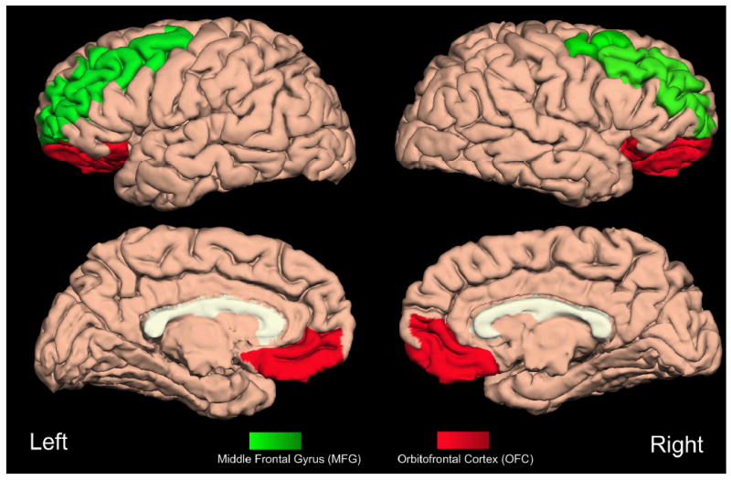
Method: Subjects included 157 patients with neurodegenerative disease. A semiautomated parcellation program (Freesurfer) was used to generate regional cortical volumes from structural MRI scans. Regions of interest (ROIs) included anterior cingulate cortex (ACC), orbitofrontal cortex (OFC), middle frontal gyrus (MFG), and inferior frontal gyrus (IFG). Socioemotional disinhibition was measured using the Neuropsychiatric Inventory. Principal component analysis including 3 tasks of executive function (EF; verbal fluency, Stroop Interference, modified Trails) was used to generate a single-factor score to represent EF.
Results: Partial correlations between ROIs, disinhibition, and EF were computed after controlling for total intracranial volume, Mini-Mental State Examination, diagnosis, age, and education. Brain regions significantly correlated with disinhibition (ACC, OFC, IFG, and temporal lobes) and EF (MFG) were entered into separate hierarchical regressions to determine which brain regions predicted disinhibition and EF. OFC was the only brain region to significantly predict disinhibition, and MFG significantly predicted EF performance. A multivariate general linear model demonstrated a significant interaction between ROIs and cognitive-behavioral functions.
Conclusions: These results support a specific association between orbitofrontal areas and behavioral management as compared with dorsolateral areas and EF.
(c) 2011 APA, all rights reserved
Figures
References
-
- Adolphs R. Cognitive neuroscience of human social behaviour. Nat Rev Neurosci. 2003;4(3):165–178. - PubMed
-
- Anderson AK, Spencer DD, Fulbright RK, Phelps EA. Contribution of the anteromedial temporal lobes to the evaluation of facial emotion. Neuropsychology. 2000;14(4):526–536. - PubMed
-
- Anderson SW, Damasio H, Jones RD, Tranel D. Wisconsin Card Sorting Test performance as a measure of frontal lobe damage. J Clin Exp Neuropsychol. 1991;13(6):909–922. - PubMed
-
- Aron AR, Robbins TW, Poldrack RA. Inhibition and the right inferior frontal cortex. Trends Cogn Sci. 2004;8(4):170–177. - PubMed
Publication types
MeSH terms
Grants and funding
- P50 AG023501/AG/NIA NIH HHS/United States
- R01 MH71940/MH/NIMH NIH HHS/United States
- R01 AG032306/AG/NIA NIH HHS/United States
- P41 RR013642/RR/NCRR NIH HHS/United States
- U54 RR021813/RR/NCRR NIH HHS/United States
- P41-RR013642/RR/NCRR NIH HHS/United States
- 5 P01-AG019724-02/AG/NIA NIH HHS/United States
- 3 P50- AG23501/AG/NIA NIH HHS/United States
- 1R01-AG022983-01/AG/NIA NIH HHS/United States
- P01 AG019724/AG/NIA NIH HHS/United States
- R01 AG022983/AG/NIA NIH HHS/United States
- R01 MH071940/MH/NIMH NIH HHS/United States