

Intrauterine inflammation, insufficient to induce parturition, still evokes fetal and neonatal brain injury
- PMID: 21382466
- PMCID: PMC3140629
- DOI: 10.1016/j.ijdevneu.2011.02.011
Intrauterine inflammation, insufficient to induce parturition, still evokes fetal and neonatal brain injury
Abstract
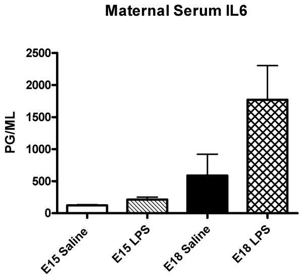
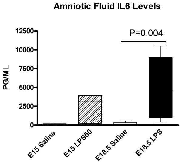
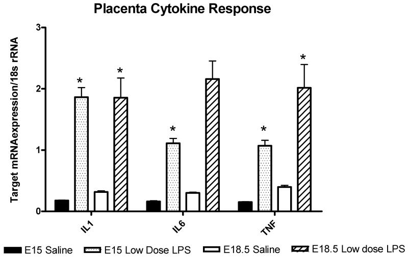
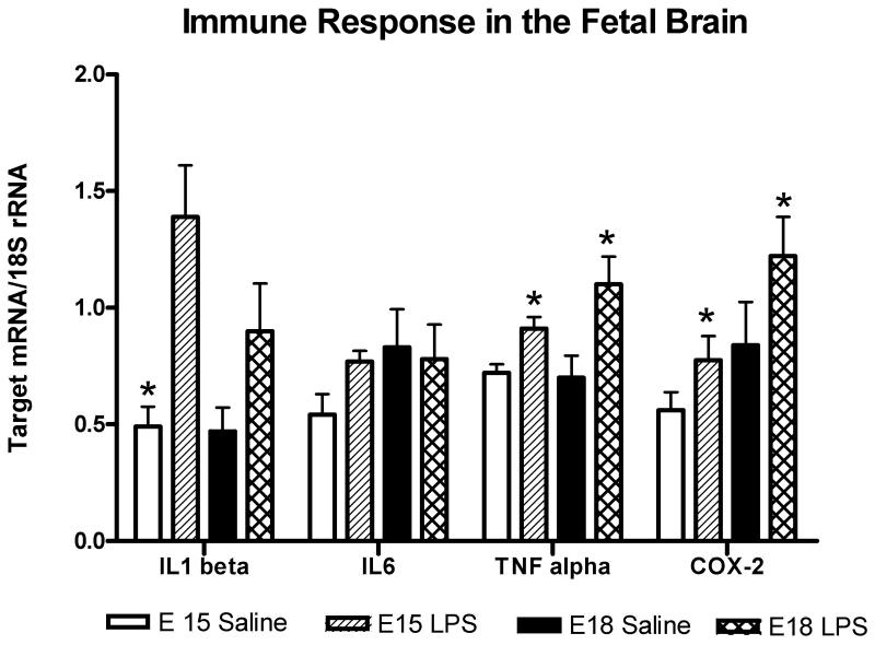
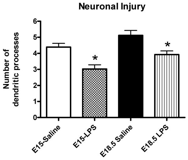
Exposure to prenatal inflammation is a known risk factor for long term neurobehavioral disorders including cerebral palsy, schizophrenia, and autism. Models of systemic inflammation during pregnancy have demonstrated an association with an immune response an adverse neurobehavioral outcomes for the exposed fetus. Yet, the most common route for an inflammatory exposure to a fetus is from intrauterine inflammation as occurs with chorioamnionitis. The aims of this study were to assess the effect of intrauterine inflammation on fetal and neonatal brain development and to determine if the gestational age of exposure altered the maternal or fetal response to inflammation. CD-1 timed pregnant mice on embryonic day 15 (E15) and E18.5 were utilized for this study. Dams were randomized to receive intrauterine infusion of lipopolysaccharide (LPS, 50 μg/dam) or normal saline. Different experimental groups were used to assess both acute and long-term outcomes. For each gestational age and each treatment group, fetal brains, amniotic fluid, maternal serum and placentas were collected 6h after intrauterine infusion. Rates of preterm birth, maternal morbidity and litter size were assessed. IL6 levels were assayed in maternal serum and amniotic fluid. An immune response was determined in the fetal brains and placentas by QPCR. Cortical cultures were performed to assess for fetal neuronal injury. Gene expression changes in postnatal day 7 brains from exposed and unexposed pups were determined. In the preterm period, low dose LPS resulted in a 30% preterm birth rate. Litter sizes were not different between the groups at either gestational age. IL6 levels were not significantly increased in maternal serum at either gestational time period. Low dose LPS increased IL6 levels in the amniotic fluid from exposed dams in the term but not preterm period. Regardless of gestational age of exposure, low dose intrauterine LPS activated an immune response in the placenta and fetal brain. Exposure to intrauterine LPS significantly decreased dendritic counts in cortical cultures from both the preterm and term period. Exposure to intrauterine inflammation altered gene expression patterns in the postnatal brain; this effect was dependent on gestational age of exposure. In conclusion, intrauterine inflammation, even in the absence of preterm parturition, can evoke fetal brain injury as evidence by alterations in cytokine expression and neuronal injury. Despite an absent or limited maternal immune response in low dose intrauterine inflammation, the immune system in the placenta is activated which is likely sufficient to induce a fetal immune response and subsequent brain injury. Changes in the fetal brain lead to changes in gene expression patterns into the neonatal period. Subclinical intrauterine inflammation can lead to fetal brain injury and is likely to be mechanistically associated with long term adverse outcomes for exposed offspring.
Copyright © 2011 ISDN. Published by Elsevier Ltd. All rights reserved.
Figures
References
-
- Abdolmaleky HM, Cheng KH, Russo A, Smith CL, Faraone SV, Wilcox M, Shafa R, Glatt SJ, Nguyen G, Ponte JF, Thiagalingam S, Tsuang MT. Hypermethylation of the reelin (RELN) promoter in the brain of schizophrenic patients: a preliminary report. Am J Med Genet B Neuropsychiatr Genet. 2005;134B:60–6. - PubMed
-
- Abecasis GR, Cherny SS, Cookson WO, Cardon LR. Merlin--rapid analysis of dense genetic maps using sparse gene flow trees. Nature Genetics. 2002;30:97–101. - PubMed
-
- Adams-Chapman I, Stoll BJ. Neonatal infection and long-term neurodevelopmental outcome in the preterm infant. Curr Opin Infect Dis. 2006;19:290–7. - PubMed
-
- Adegbola AA, Gonzales ML, Chess A, LaSalle JM, Cox GF. A novel hypomorphic MECP2 point mutation is associated with a neuropsychiatric phenotype. Hum Genet. 2009;124:615–23. - PubMed
