Review
. 2011 Jun;29(4):423-40.
doi: 10.1016/j.ijdevneu.2011.02.012. Epub 2011 Mar 5.

The developing oligodendrocyte: key cellular target in brain injury in the premature infant

Affiliations
Review

The developing oligodendrocyte: key cellular target in brain injury in the premature infant

Joseph J Volpe et al. Int J Dev Neurosci. 2011 Jun.

Abstract

Brain injury in the premature infant, a problem of enormous importance, is associated with a high risk of neurodevelopmental disability. The major type of injury involves cerebral white matter and the principal cellular target is the developing oligodendrocyte. The specific phase of the oligodendroglial lineage affected has been defined from study of both human brain and experimental models. This premyelinating cell (pre-OL) is vulnerable because of a series of maturation-dependent events. The pathogenesis of pre-OL injury relates to operation of two upstream mechanisms, hypoxia-ischemia and systemic infection/inflammation, both of which are common occurrences in premature infants. The focus of this review and of our research over the past 15-20 years has been the cellular and molecular bases for the maturation-dependent vulnerability of the pre-OL to the action of the two upstream mechanisms. Three downstream mechanisms have been identified, i.e., microglial activation, excitotoxicity and free radical attack. The work in both experimental models and human brain has identified a remarkable confluence of maturation-dependent factors that render the pre-OL so exquisitely vulnerable to these downstream mechanisms. Most importantly, elucidation of these factors has led to delineation of a series of potential therapeutic interventions, which in experimental models show marked protective properties. The critical next step, i.e., clinical trials in the living infant, is now on the horizon.


Figures

Fig. 1
Fig. 1
Periventricular leukomalacia. Coronal section of cerebrum (H+E stain) in a premature infant who died several weeks after a cardiac arrest. Note the two components of the lesion (i.e., deep focal areas of cystic necrosis and more diffuse cerebral white matter injury). See text for details. (Courtesy of Dr. Hannah C. Kinney).
Fig. 2
Fig. 2
Progression of the oligodendroglial lineage (OL) through the four major stages. The predominant form in cerebral white matter of the premature infant is the 04-positive pre-oligodendrocyte. See text for details. (From Back SA, Volpe JJ, Ment Retard Dev Disabil Res Rev 3: 96-107, 1997). Note that in this review the abbreviation pre-OL includes both premyelinating OL forms, i.e., the 04-positive pre-oligodendrocyte and the 01-positive immature oligtodendrocytes.
Fig. 3
Fig. 3
Cystic (A) and noncystic (B) periventricular leukomalacia (PVL) – schematic diagrams. A. Cystic PVL is characterized by macroscopic (several mm or more) focal necrotic lesions that become cystic and by diffuse astrogliosis and pre-OL injury. B. Noncystic PVL is characterized by focal necrotic lesions that are microscopic and evolve principally to small glial scars rather than cysts.
Fig. 4
Fig. 4
04 immunostaining of pre-OLs in control (A) and PVL (B) cases. Pre-OLs in control cases (A) are multipolar with multiple discrete processes (arrow), whereas in PVL (B) some pre-OLs lack processes (arrows). (From Billiards SS, et al, Brain Pathol 18: 153-163, 2008).
Fig. 5
Fig. 5
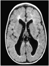
Axial MRI (FLAIR) of cerebrum in a 20-month-old infant who was born prematurely and had PVL. Note the marked paucity of cerebral white matter and, as a consequence, enlarged lateral ventricles. The increased signal intensity in white matter is caused by astrogliosis. (Courtesy of Dr. Linda deVries.)
Fig. 6
Fig. 6
Pathogenesis of PVL. The two major upstream mechanisms (pink) are ischemia and systemic infection/inflammation, activating three major downstream mechanisms (blue), microglial activation, glutamate excitotoxicity and ultimately, free radical attack. See text for details.
Fig. 7
Fig. 7
Oxidative (upper panel) and nitrative (lower panel) injury in PVL. Hydroxynonenal (HNE) staining (upper panel), a marker for attack by reactive oxygen species, colocalizes (yellow) with a marker of pre-OLs (04) in the diffuse component of PVL. Nitrotyrosine (NT) staining, (lower panel), a marker for attack by reactive nitrogen species, colocalizes (yellow) with pre-OLs (04) in the diffuse component of PVL as well. (From Haynes RL, et al, J Neuropathol Exp Neurol 62: 441-450, 2003).
Fig. 8
Fig. 8
Regression curves of analyses of antioxidant enzyme expressions (Western blots) in cerebral white matter from midgestation to the first postnatal weeks, relative to the adult standard (100% value). Immunocytochemical studies showed similar temporal changes in pre-OLs. The peak period for occurrence of PVL is shown by the black bar. (From Folkerth RD, et al, J Neuropathol Exp Neurol 63: 990-999, 2004).
Fig. 9
Fig. 9
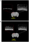
Protection from hypoxic-ischemic selective cerebral white matter injury in the immature rat (P7) by systemic administration of topiramate. In 9A, the top panels show a pronounced deficit in myelin-basic protein staining in cerebral white matter at P11, 4 days after hypoxemia-ischemia at P7. Compare in 9A the hemispheres ipsilateral (left) and contralateral (right) to the carotid ligation. (In separate experiments marked loss of pre-OLs was demonstrated in the ipsilateral hemisphere in the 24 hours after the insult.) In 9B, topiramate was administered intraperitoneally over the 48 hours following the insult, beginning immediately after termination of the insult. A marked protective effect is apparent. (From Follett, P, et al, J Neurosci 24: 4412-4420, 2004).
Fig. 10
Fig. 10
Protection from hypoxic-ischemic selective cerebral white matter injury in the immature (P7) rat by systemic administration of the NMDA receptor blocker, memantine. The paradigm is similar to that described in the legend to Fig. 9, except that memantine rather than topiramate was administered systemically after termination of the insult. A (ipsilateral to carotid ligation) and B (contralateral) were vehicle-treated and C (ipsilateral) and D (contralateral) were memantine-treated. Note the pronounced protective effect of memantine (compare A to C). (From Manning SM, et al, J Neurosci 28: 6670-6678, 2008.)
Fig. 11
Fig. 11
Potential differential effects and temporal aspects of excitotoxicity to developing oligodendrocytes. The intact cell (top) has AMPA receptors primarily on the cell soma and NMDA receptors primarily on the cell processes. Initially with excess extracellular glutamate, activation of NMDA receptors could lead to loss of cell processes, and if excitotoxicity continues, to activation of AMPA receptors and cell death. Either event could lead to impaired myelination (solid arrows) and potentially also to axonal disturbance (dotted lines).
Fig. 12
Fig. 12
Microglia and innate immune mechanisms in pre-OL injury. Microglia may act as a convergence point for both upstream mechanisms in PVL, i.e., systemic infection/inflammation and hypoxia-ischemia, and innate immunity is likely involved in both microglial mechanisms. Thus, pathogen-associated molecular patterns (PAMPs) and danger-associated molecular patterns (DAMPs), respectively lead to microglial activation and resulting release of products, especially reactive oxygen and nitrogen species (ROS/RNS) and cytokines, that result in pre-OL injury. See text for details.
Fig. 13
Fig. 13
Potential mechanism by which endogenous TLR ligands, specifically hyaluronan and TLR2, may lead to inhibition of pre-OL differentiation to mature myelin-producing OLs. See text for details.
Fig. 14
Fig. 14
Interventions for prevention of injury to developing OLs resulting from hypoxic-ischemic and inflammatory insults. See text for details.

Republished in

References

    1. Aarnoudse-Moens CS, Weisglas-Kuperus N, van Goudoever JB, Oosterlaan J. Meta-analysis of neurobehavioral outcomes in very preterm and/or very low birth weight children. Pediatrics. 2009;124:717–728. - PubMed
    1. Agresti C, D’Urso D, Levi G. Reversible inhibitory effects of interferon-γ- and tumour necrosis factor-α on oligodendroglial lineage cell proliferation and differentiation in vitro. Eur J Neurosci. 1996;8:1106–1116. - PubMed
    1. Alberdi E, Sanchez-Gomez MV, Marino A, Matute C. Ca(2+) influx through AMPA or kainate receptors alone is sufficient to initiate excitotoxicity in cultured oligodendrocytes. Neurobiol Dis. 2002;2:234–243. - PubMed
    1. Albrecht P, Lewerenz J, Dittmer S, Noack R, Maher P, Methner A. Mechanisms of oxidative glutamate toxicity: the glutamate/cystine antiporter system xc- as a neuroprotective drug target. CNS Neurol Disord Drug Targets. 2010;9:373–382. - PubMed
    1. Andiman SE, Haynes RL, Trachtenberg FL, Billiards SS, Folkerth RD, Volpe JJ, Kinney HC. The cerebral cortex overlying periventricular leukomalacia: Analysis of pyramidal neurons. Brain Pathol. 2010;20:803–814. - PMC - PubMed

Publication types

MeSH terms