. 2011 May 15;50(10):1252-62.
doi: 10.1016/j.freeradbiomed.2011.02.031. Epub 2011 Mar 5.

Caveolin-1 mediates Fas-BID signaling in hyperoxia-induced apoptosis

Affiliations

Caveolin-1 mediates Fas-BID signaling in hyperoxia-induced apoptosis

Meng Zhang et al. Free Radic Biol Med. .

Abstract

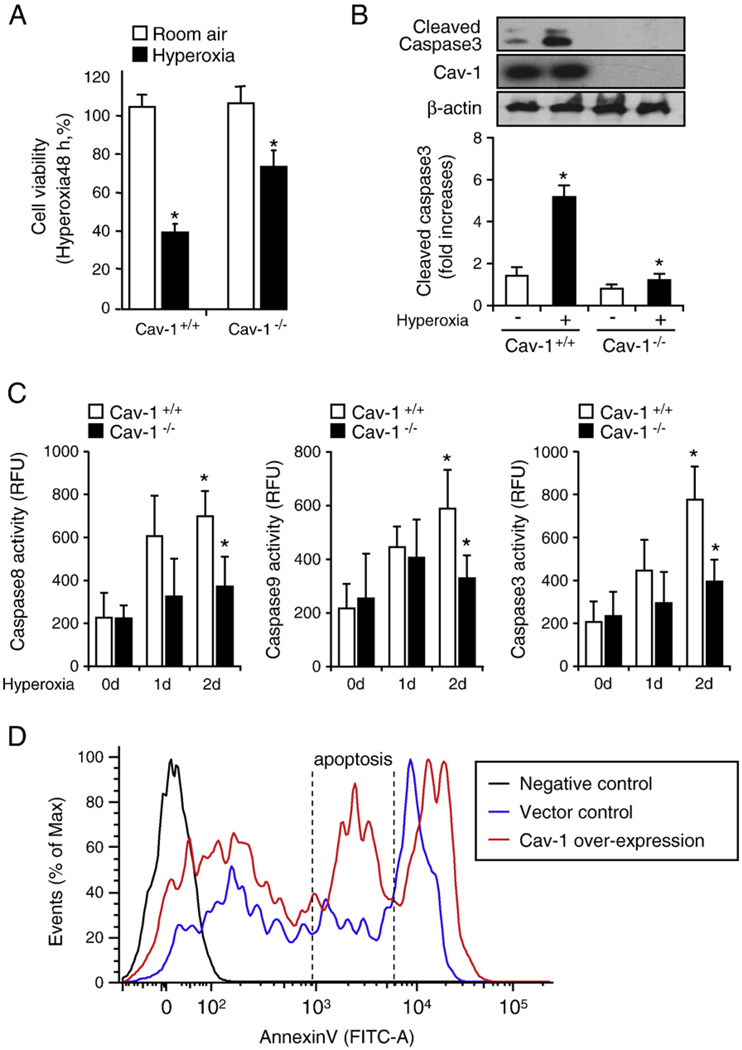
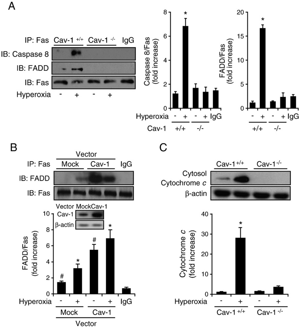
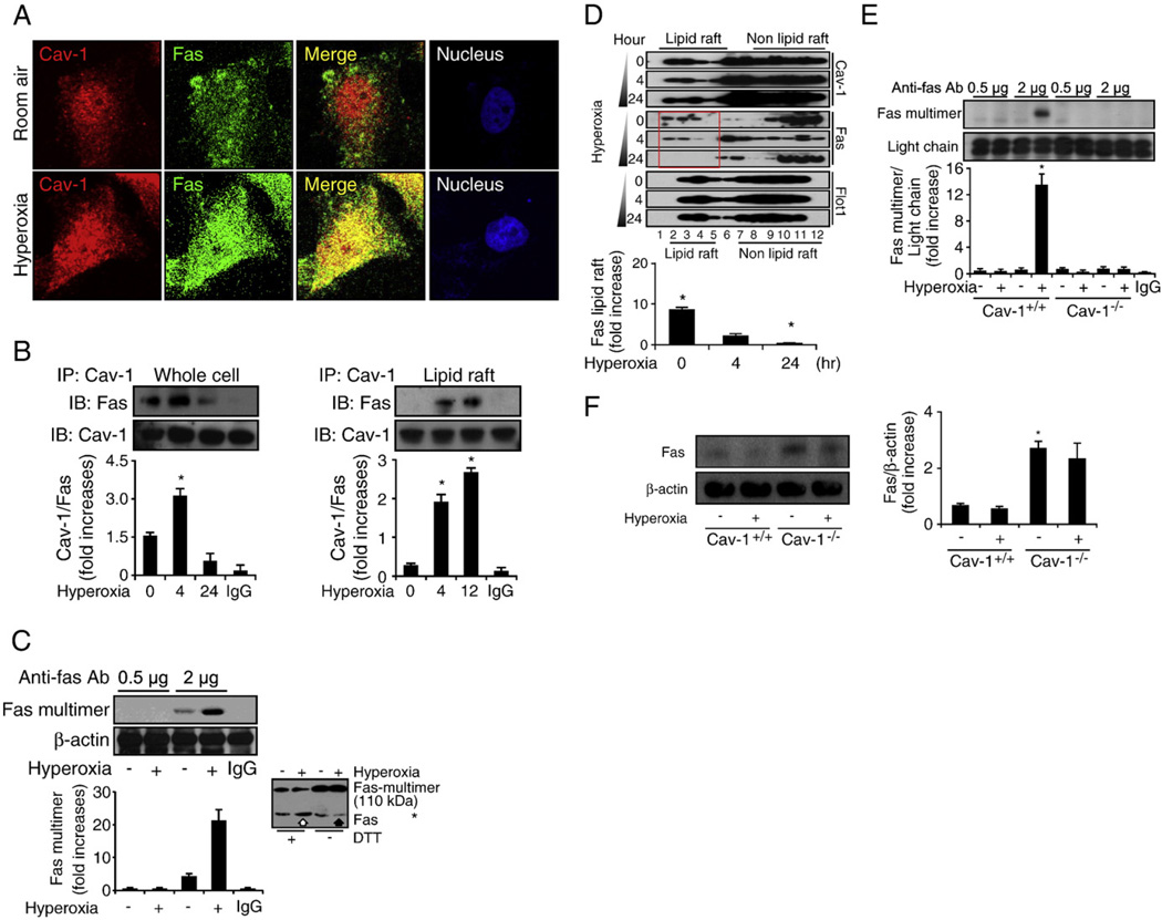
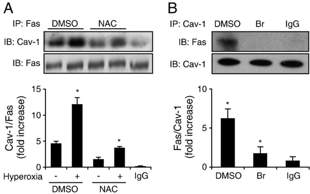
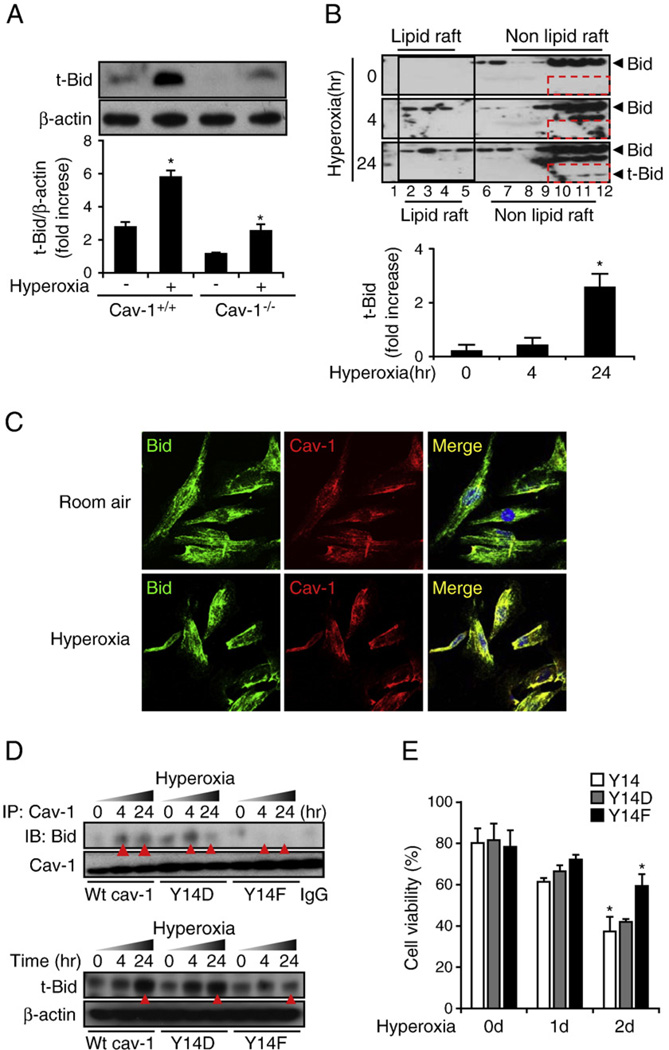
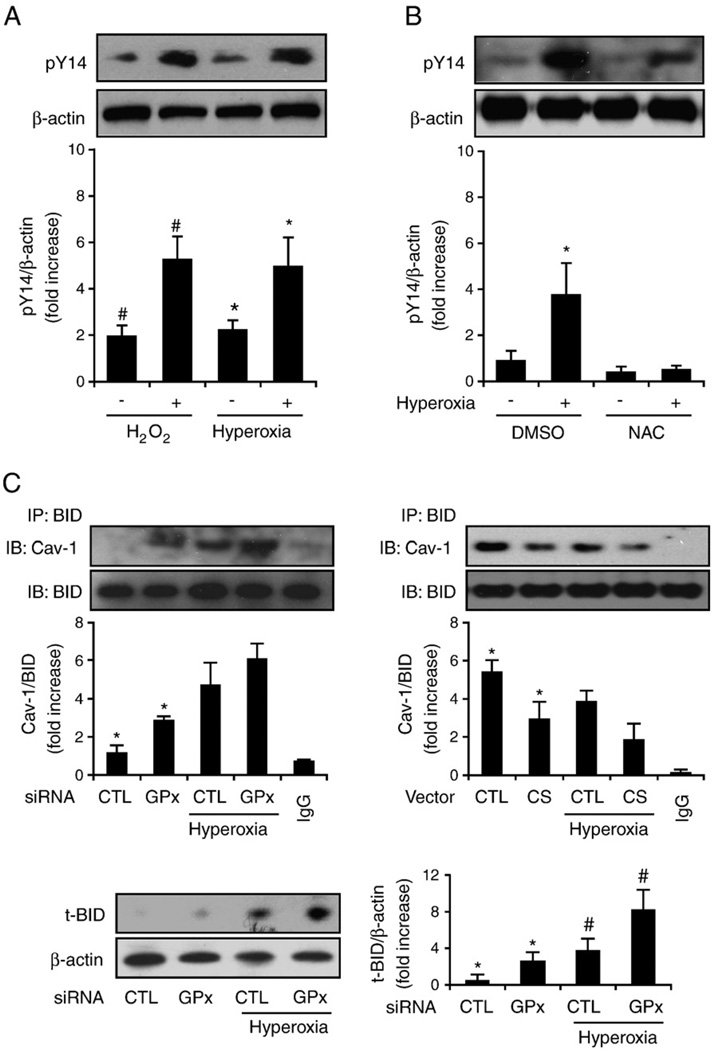
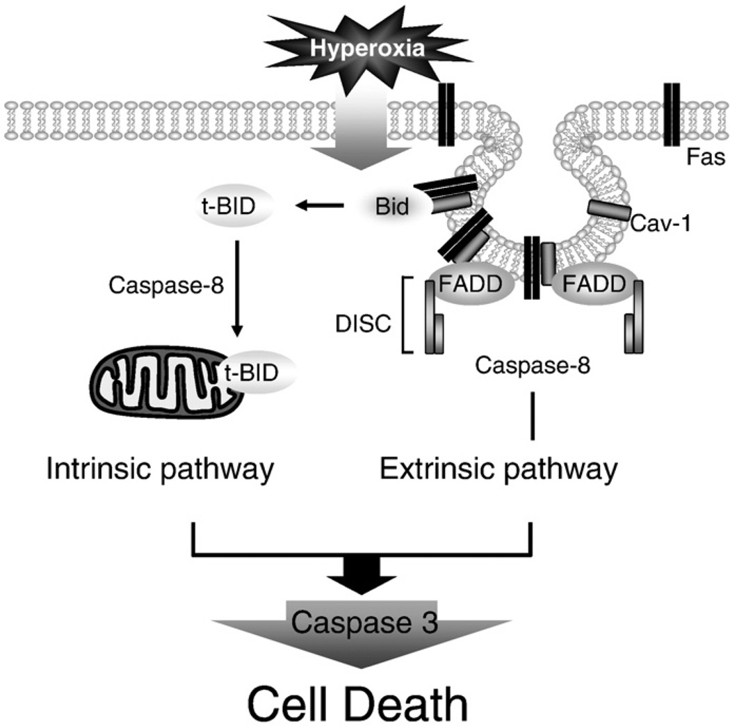
Fas-mediated apoptosis is a crucial cellular event. Fas, the Fas-associated death domain, and caspase 8 form the death-inducing signaling complex (DISC). Activated caspase 8 mediates the extrinsic pathways and cleaves cytosolic BID. Truncated BID (tBID) translocates to the mitochondria, facilitates the release of cytochrome c, and activates the intrinsic pathways. However, the mechanism causing these DISC components to aggregate and form the complex remains unclear. We found that Cav-1 regulated Fas signaling and mediated the communication between extrinsic and intrinsic pathways. Shortly after hyperoxia (4 h), the colocalization and interaction of Cav-1 and Fas increased, followed by Fas multimer and DISC formation. Deletion of Cav-1 (Cav-1-/-) disrupted DISC formation. Further, Cav-1 interacted with BID. Mutation of Cav-1 Y14 tyrosine to phenylalanine (Y14F) disrupted the hyperoxia-induced interaction between BID and Cav-1 and subsequently yielded a decreased level of tBID and resistance to hyperoxia-induced apoptosis. The reactive oxygen species (ROS) scavenger N-acetylcysteine decreased the Cav-1-Fas interaction. Deletion of glutathione peroxidase-2 using siRNA aggravated the BID-Cav-1 interaction and tBID formation. Taken together, these results indicate that Cav-1 regulates hyperoxia/ROS-induced apoptosis through interactions with Fas and BID, probably via Fas palmitoylation and Cav-1 Y14 phosphorylation, respectively.


Figures

Fig. 1
Fig. 1
Deletion of Cav-1 protected lung epithelial cells against hyperoxia-induced apoptosis via the regulation of apoptotic pathways. Primary mouse lung epithelial cells were used in these experiments. Cells were exposed to room air (i.e., 20.8% oxygen + 78.08% nitrogen) or hyperoxia (95% oxygen + 5% balanced nitrogen) conditions. At designated times (i.e., after 4, 24, and 48 h), cells were collected and subjected to cell survival assays, namely, Western blot analysis or caspase activity assay. (A) The deletion of Cav-1 protected the primary mouse lung epithelial cells from hyperoxia-induced cell death. Primary mouse lung epithelial cells were isolated from either wild-type C57BL/6 mice (Cav-1+/+) or Cav-1−/− mice. The cells were then exposed to room air or hyperoxia conditions. After 48 h, cell viability was determined, as described under Materials and methods. (B) The deletion of Cav-1 protected the primary mouse lung epithelial cells against hyperoxia-induced activation of caspase 3. The epithelial cells were isolated from either wild-type C57BL/6 mice or Cav-1−/− mice. The cells were then exposed to hyperoxia conditions (48 h), after which cell lysates were obtained and subjected to Western blot analysis. (C) The deletion of Cav-1 protected the primary mouse lung epithelial cells against hyperoxia-induced caspase 3, 8, and 9 activation. Cells were exposed to room air, 24 h hyperoxia, or 48 h hyperoxia conditions. After exposure, cell lysates were obtained and caspase activities were measured using assays as described previously [40,41]. (D) To confirm the proapoptotic effect of Cav-1, we overexpressed Cav-1 or empty vectors in Beas-2B cells. Cell death was analyzed using FACS. Live cells were directly stained with annexin V–FITC and Sytox green dye. The Sytox green dye is impermeative to live cells and apoptotic cells, but stains necrotic cells with intense green fluorescence. After staining, apoptotic cells show green fluorescence, whereas dead cells show a higher level of green fluorescence and live cells show little or much lower levels of fluorescence. Three cell populations were observed, including (1) live cells, (2) apoptotic cells, and (3) necrotic cells. All experiments presented were repeated using three independent assays with similar results. *P<0.05.
Fig. 2
Fig. 2
Cav-1 facilitated the hyperoxia-induced Fas–FADD/caspase 8 interaction in the absence of the Fas ligand. Primary mouse lung epithelial cells and Beas-2B human epithelial cells were used in these experiments. Cells were exposed to room air (i.e., 20.8% oxygen + 78.08% nitrogen) or hyperoxia (95% oxygen + 5% balanced nitrogen) conditions. At designated times (i.e., after 4, 24, or 48 h), the cells were collected and subjected to co-IP assay or Western blot analysis. DISC formation was determined by detecting the interaction of Fas–FADD/caspase 8. (A) The deletion of Cav-1 disrupted hyperoxia-induced DISC formation. Primary mouse lung epithelial cells from either wild-type C57BL/6 mice (Cav-1+/+) or Cav-1−/− mice were exposed to room air or hyperoxia (4 h) conditions. Co-IP assays between Fas and FADD and Fas and caspase 8 were performed as described previously. (B) Overexpressing Cav-1 facilitated Fas–FADD interaction after hyperoxia. Beas-2B cells were used in these experiments and infected with adeno-lacZ (control) and adeno-Cav-1, as described previously [41]. Cells were then exposed to hyperoxia (4 h), and co-IP assays were performed between Fas and FADD. (C) Deletion of Cav-1 prevented the release of cytochrome c after hyperoxia. Primary mouse lung epithelial cells from either wild-type C57BL/6 mice or Cav-1−/− mice were exposed to hyperoxia. After 24 h, the level of cytosol cytochrome c was determined by Western blot analysis, as described previously [28]. All experiments presented were repeated using three independent assays with similar results. *P<0.05,#P<0.01.
Fig. 3
Fig. 3
Cav-1 directly interacts with Fas and facilitates Fas–Fas multimerization after hyperoxia. Primary mouse lung epithelial cells and Beas-2B cells were used in these experiments and treated with RA or hyperoxia conditions for the designated times. (A) The colocalization between Cav-1 and Fas after hyperoxia (4 h). Beas-2B cells were exposed to RA or hyperoxia, as described previously. After 4 h, the cells were stained with anti-Fas, anti-Cav-1, and DAPI and then subjected to confocal microscopy. (B) Cav-1–Fas interaction was determined by co-IP assays. Beas-2B cells were exposed to RA or hyperoxia conditions, as indicated. On the left, whole-cell lysate was used in the co-IP assays. On the right, isolated lipid rafts were used to perform co-IP. Reverse co-IP was performed with similar results (i.e., IP, Fas; IB, Cav-1). (C) Hyperoxia-induced Fas–Fas multimerization in Beas-2B cells. Cells were exposed to hyperoxia (4 h), followed by Fas multimerization assays, as described previously [47]. Briefly, a limited amount of rabbit polyclonal anti-Fas antibody was used to precipitate Fas multimer. After fractioning by SDS–PAGE, mouse monoclonal anti-Fas antibody was used as the primary antibody to detect the precipitated Fas multimer. Using a limited amount of antibodies (0.5–2 µg), a visible difference was observed for the amount of precipitated Fas multimers between RA-treated and hyperoxia-treated cells. When an excess amount of antibodies (15–20 µg) was used to precipitate the Fas multimer, there was no significant difference between any of the groups (not shown). (D) Hyperoxia induced Fas trafficking from the lipid rafts portion to the non-lipid rafts portion, which was a time-dependent process. Beas-2B cells were exposed to RA or hyperoxia conditions. Lipid rafts were isolated as described under Materials and methods. Western blot analysis was used to detect Fas and Cav-1. (E) The deletion of Cav-1 prevented the formation of the Fas multimer after hyperoxia. Primary wild-type mouse lung epithelial cells (Cav-1+/+) versus cells isolated from Cav-1−/− mice were used, and Fas multimerization was determined as described previously. (F) The deletion of Cav-1 caused the Fas expression to be up-regulated in both the absence and the presence of hyperoxia. Primary mouse lung epithelial cells were isolated, and cells were exposed to RA or hyperoxia (24 h). The protein level of Fas was determined by using Western blot analysis. For each panel, three independent assays are represented. *P<0.05, #P<0.01.
Fig. 4
Fig. 4
Cav-1 interacts with Fas after hyperoxia, partially via ROS and palmitoylation. (A) Blocking ROS with NAC (30 nM) partially eliminated Cav-1–Fas interaction. Beas-2B cells were pretreated with an NAC or PBS control, and then after 30 min, the cells were exposed to hyperoxia (4 h). Cell lysates were then collected, and co-IP assays were performed to determine Cav-1–Fas interactions. (B) Cav-1–Fas interaction after hyperoxia is mediated by palmitoylation. Beas-2B cells were pretreated with 2-Br (1 µM), and then after 30 min, the cells were exposed to hyperoxia (4 h). The Cav-1–Fas interaction was determined by using co-IP assays, as described previously. All blots represent three repeats. *P<0.05.
Fig. 5
Fig. 5
Cav-1 interacts with BID and regulates the hyperoxia-induced intrinsic and extrinsic apoptotic pathways. Primary mouse lung epithelial cells and Beas-2B human bronchial epithelial cells were used in this study. (A) The deletion of Cav-1 prevented the formation of truncated BID. Primary wild-type mouse lung epithelial cells (Cav-1+/+) versus cells isolated from Cav-1−/− mice were used. Cells were exposed to hyperoxia (24 h), and the tBID level was detected. (B) Hyperoxia induced BID trafficking and tBID formation in Beas-2B cells. The lipid raft was isolated, as described previously. BID was then determined by using Western blot analysis. Using goat polyclonal anti-BID (Santa Cruz Biotechnology), both the 22- and the 15-kDa segments may be detected. Hyperoxia induced BID trafficking from the nonlipid portion to the lipid portion of the rafts. (C) The colocalization between Cav-1 and BID in the absence or presence of hyperoxia (4 h). Beas-2B cells were stained with anti-Cav-1, anti-BID, and DAPI. The merged images are shown (right), representing the colocalization of Cav-1 and BID in the presence and absence of hyperoxia. (D) The mutation of Cav-1 phosphorylated tyrosine Y14 to Y14F disrupted the interaction between Cav-1 and BID. Two mutated clones (Y14F and Y14D), in addition to the wild-type Y14, were transfected into Beas-2B cells. The cells were then exposed to hyperoxia for 4 or 24 h. The Cav-1–BID interaction was determined by using co-IP assays. The tBID level was determined by using Western blot analysis. (E) Beas-2B cells that were transfected with Cav-1 Y14F, but not the wild-type Y14 or Y14D, show cytoprotective effects against hyperoxia-induced cell death. As described previously, after transfection with Y14, Y14D, or Y14F, Beas-2B cells were exposed to hyperoxia. After a designated time-frame, cell viability was determined as described under Materials and methods. All the above experiments represent a minimum of three repeats. *P<0.05.
Fig. 6
Fig. 6
Hyperoxia induces Cav-1 phosphorylation via ROS. Beas-2B human bronchial epithelial cells and primary mouse lung epithelial cells were used in these experiments. (A) Hyperoxia and H2O2 induce Cav-1 phosphorylation. Primary lung epithelial cells derived from C57BL/6 mice were incubated with 90 µM H2O2 for 20 min, exposed to hyperoxia (4 h), or left untreated. Cell lysates were collected and analyzed by Western blotting and were blotted with anti-PY14Cav-1. (B) NAC blocks hyperoxia-induced Cav-1 Y14 phosphorylation. Primary lung epithelial cells were pretreated with NAC (30 nM), which was followed by exposure to hyperoxia (4 h). Western blot analysis was used to determine Y14 phosphorylation. Similar results were found in Beas-2B cells. (C) Beas-2B cells were transfected with GPX2 siRNA or control siRNA (left) or with catalase overexpression clones or control vectors (right). After 24 h, cells were exposed to hyperoxia (4 h). Coimmunoprecipitation of Cav-1 and BID was determined. Cell lysates were incubated with anti-BID antibody overnight at 4 °C and then coupled with protein A/G agarose beads. The Cav-1 protein, which coimmunoprecipitated with BID, was detected by Western blotting. Incubation of the same amount of lysate with rabbit IgG was used as a negative control. A representative of at least three experiments is shown. IP, immunoprecipitation; IB, immunoblot. (C, bottom) Cells transfected with GPX2 siRNA have elevated tBID. Beas-2B cells were transfected with GPX2 siRNA or control siRNA as above. tBID was determined by Western blot analysis. *P<0.05, #P<0.05.
Fig. 7
Fig. 7
Schematic illustration of the mechanisms by which Cav-1 regulates hyperoxia-induced extrinsic and intrinsic apoptotic pathways in lung epithelial cells. Hyperoxia induces Fas/Cav-1 palmitoylation and hence induces Fas–Cav-1 interaction. By interacting with Fas, Cav-1 facilitates Fas multimerization and initiates DISC formation, which subsequently activates caspase 8. Meanwhile, hyperoxia induces Cav-1 Y14 phosphorylation, which then induces Cav-1–BID interaction. The interaction between Cav-1 and BID via Y14 phosphorylation increases the proximity of BID to activated caspase 8. Therefore, Cav-1 also facilitates the truncation of BID to tBID by caspase 8. tBID translocates to the mitochondria and mediates the release of mitochondrial cytochrome c, which induces intrinsic apoptotic pathways, resulting in elevated caspase 9 activity.

Similar articles

Cited by

References

    1. Ashkenazi A, Dixit VM. Death receptors: signaling and modulation. Science. 1998;281:305–1308. - PubMed
    1. Takahashi T, Tanaka M, Inazawa J, Abe T, Suda T, Nagata S. Human Fas ligand: gene structure, chromosomal location and species specificity. Int. Immunol. 1994;6:1567–1574. - PubMed
    1. Ogasawara J, et al. Lethal effect of the anti-Fas antibody in mice. Nature. 1993;364:806–809. - PubMed
    1. Jones RA, et al. Fas-mediated apoptosis in mouse hepatocytes involves the processing and activation of caspases. Hepatology. 1998;27:1632–1642. - PubMed
    1. Algeciras-Schimnich A, Shen L, Barnhart BC, Murmann AE, Burkhardt JK, Peter ME. Molecular ordering of the initial signaling events of CD95. Mol. Cell. Biol. 2002;22:207–220. - PMC - PubMed

Publication types