Review
. 2011 Oct 15;179(1):34-42.
doi: 10.1016/j.resp.2011.03.002. Epub 2011 Mar 5.

Development of synaptic transmission to respiratory motoneurons

Affiliations
Review

Development of synaptic transmission to respiratory motoneurons

Albert J Berger. Respir Physiol Neurobiol. .

Abstract

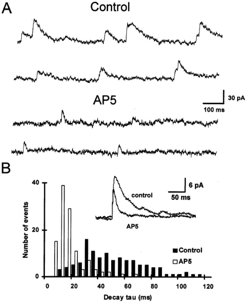
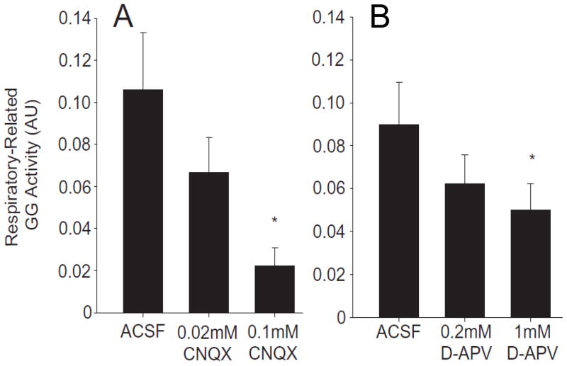
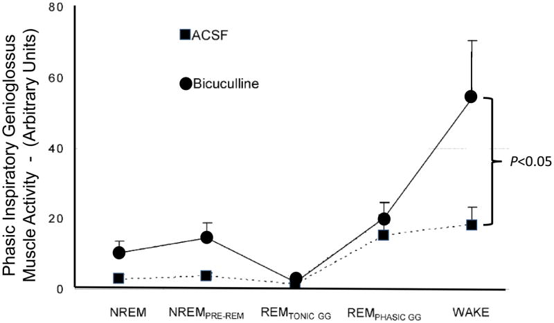
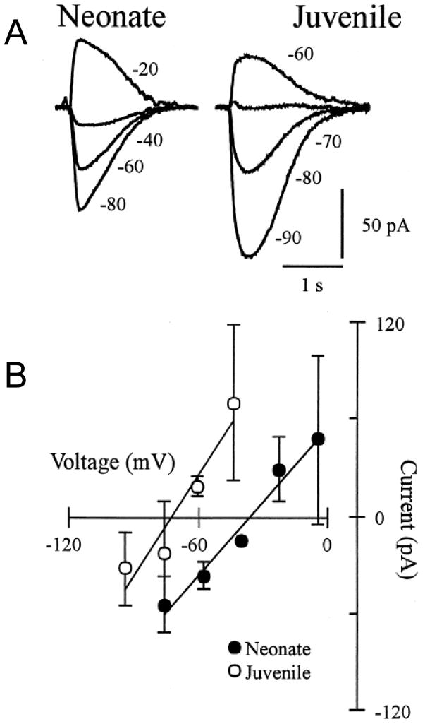
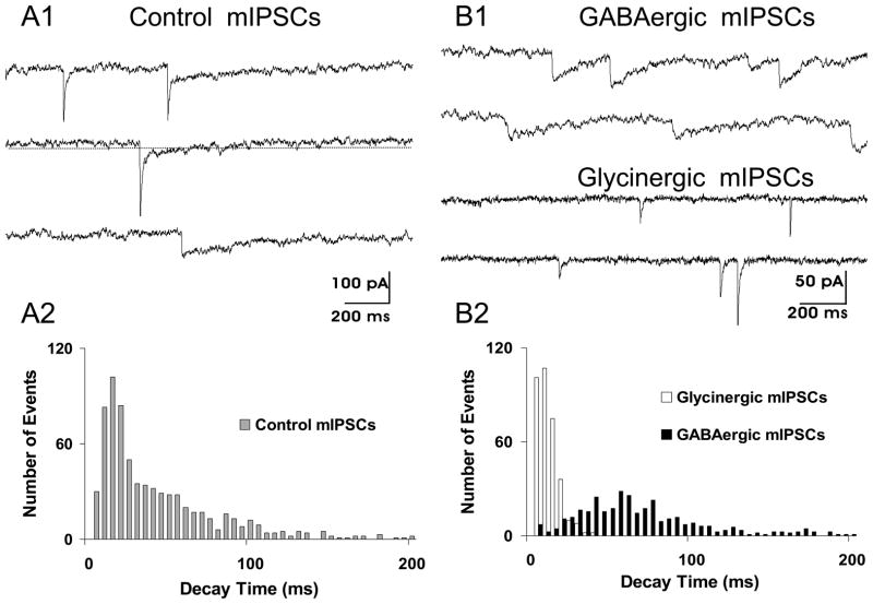
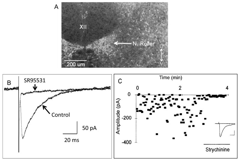
Respiratory motoneurons provide the exclusive drive to respiratory muscles and therefore are a key relay between brainstem neural circuits that generate respiratory rhythm and respiratory muscles that control moment of gases into and out of the airways and lungs. This review is focused on postnatal development of fast ionotropic synaptic transmission to respiratory motoneurons, with a focus on hypoglossal motoneurons (HMs). Glutamatergic synaptic transmission to HMs involves activation of both non-NMDA and NMDA receptors and during the postnatal period co-activation of these receptors located at the same synapse may occur. Further, the relative role of each receptor type in inspiratory-phase motoneuron depolarization is dependent on the type of preparation used (in vitro versus in vivo; neonatal versus adult). Respiratory motoneurons receive both glycinergic and GABAergic inhibitory synaptic inputs. During inspiration phrenic and HMs receive concurrent excitatory and inhibitory synaptic inputs. During postnatal development in HMs GABAergic and glycinergic synaptic inputs have slow kinetics and are depolarizing and with postnatal development they become faster and hyperpolarizing. Additionally shunting inhibition may play an important role in synaptic processing by respiratory motoneurons.


Figures

Fig. 1
Fig. 1
NMDA and non-NMDA glutamatergic receptors are co-localized in the postsynaptic membrane of hypoglossal motoneurons. This is shown by the presence of spontaneous miniature excitatory postsynaptic currents (mEPSCs) that have dual temporal components. (A) When recorded in the presence of bicuculline to block GABAA-receptors, strychnine to block glycine receptors and TTX to block action potential generated synaptic events, the majority of mEPSCs show slow decay kinetics (Control – top two traces). With the addition of AP5 to block NMDA receptors the slow decaying component is abolished leaving only a fast decaying component due that is due to activation of non-NMDA receptors (AP5 - lower two traces). (B) Distribution of mEPSC decay time constants before and after application of AP5. Inset: Average of mEPSCs before and after addition of AP5. Data show that the AP5 sensitive component of the mEPSC has a significantly longer decay time constant. Data from HMs voltage clamped at +50 mV to remove the voltage-dependent Mg block of the NMDA receptor. (Adapted from O’Brien et al., 1997).
Fig. 2
Fig. 2
In the adult anesthetized rat both non-NMDA and NMDA receptors in the hypoglossal motor nucleus contribute to the excitatory synaptic I-phase drive to HMs. Shown are data for respiratory-related genioglossus muscle activity (GG activity) in response to microdialysis perfusion of the non-NMDA receptor antagonist CNQX and separately of the NMDA receptor antagonist D-APV (also known as AP5) into the hypoglossal motor nucleus. (A) Shows the dose-dependent reduction of respiratory-related activity with CNQX. (B) Shows the dose-dependent reduction with D-APV. Data reported at means ± SEM and * indicates a P< 0.05 level of significance compared to microdialysis perfusion of the artificial cerebrospinal fluid (ACSF) control solution. (Adapted from Steenland et al., 2006).
Fig. 3
Fig. 3
Microdialysis of bicuculline into the hypoglossal motor nucleus blocks GABAA-receptors and significantly increases inspiratory-phase related hypoglossal motoneuron activity in the awake rat. Shown are group data assessing changes in respiratory-related genioglossus muscle activity (GG activity) across sleep-wake states during microdialysis perfusion of artificial cerebrospinal fluid (ACSF) control solution and bicuculline into the hypoglossal motor nucleus during room-air breathing. Data reported at means ± SEM. (Adapted from Morrison et al., 2003b).
Fig. 4
Fig. 4
The reversal potential of glycine-receptor-mediated currents recorded in HMs becomes more hyperpolarized with postnatal development. (A) Local glycine application onto a voltage-clamped HM shows the reversal potential from a neonate (P2) and a juvenile (P15) rat. Various holding potentials indicated in mV. (B) Neonate and juvenile group data for the current-voltage relationship of the glycine-evoked responses. Results show the hyperpolarizing shift in the glycine-evoked response with postnatal development. All recordings were done using the gramicidin perforated-patch technique so as not to disturb the native intracellular chloride concentration. (Adapted from Singer et al., 1998).
Fig. 5
Fig. 5
Co-transmission of GABA and glycine to a HM is shown by the presence of dual component spontaneous miniature inhibitory postsynaptic currents (mIPSCs). (A1) Control recording show the presence of three types of mIPSCs based on their temporal kinetics. Slow decaying GABAergic mIPSCs, fast decaying glycinergic mIPSCs and dual component mIPSCs, having both fast and slow decaying components. Presumably the dual component mIPSCs are due to co-release of both GABA and glycine from a single presynaptic vesicle. (A2) Distribution of mIPSC decay times in control conditions showing that the distribution is skewed toward longer decay times. (B1) In the presence of strychnine to block glycine receptors, isolated slow decaying GABAergic mIPSCs are recorded (top two traces). In the presence of bicuculline to block GABAA receptors, isolated fast decaying glycinergic mIPSCs are recorded (lower two traces). (B2) Distribution of mIPSC decay times of GABAergic and glycinergic mIPSCs shows two unique distributions (P<0.001) based on Kolmogorov-Smirnoff statistical testing of these distributions. (Adapted from O’Brien and Berger, 1999).
Fig. 6
Fig. 6
The Nucleus of Roller located just ventrolateral to the hypoglossal motor nucleus contains GABAergic and glycinergic neurons that monosynaptically inhibit HMs. (A) A confocal fluorescent photomicrograph of a transverse section of the medulla from a GAD67-GFP knock-in mouse showing the high density of GABAergic cells in the Nucleus of Roller, but an absence of GFP containing cells within the hypoglossal motor nucleus (XII). (B) Local electrical stimulation of the Nucleus of Roller produces GABAA receptor-mediated currents in a HM. Data shown are average traces of GABAA receptor-mediated evoked IPSCs before and during bath application of SR95531 (also known as gabazine) to block GABAA receptors. Application of SR95531 almost completely blocked the evoked response. (C) Local electrical stimulation of the Nucleus of Roller also can produce glycine-receptor-mediated currents in a HM. Data points are the amplitude of single evoked IPSCs plotted against time. Strychnine applied during the time indicated almost completely abolished the evoked glycinergic IPSCs. Inset: Shows average traces before and during application of strychnine. Calibration bars indicate 50 pA and 20 ms. (A and B adapted from van Brederode et al., 2011; C adapted from Umemiya and Berger, 1995).

References

    1. Aldes LD, Chronister RB, Marco LA. Distribution of glutamic acid decarboxylase and gamma-aminobutyric acid in the hypoglossal nucleus in the rat. J Neurosci Res. 1988;19:343–348. - PubMed
    1. Altmann H, ten Bruggencate G, Sonnhof U. Differential strength of action of glycine and GABA in hypoglossus nucleus. Pflugers Arch. 1972;331:90–94. - PubMed
    1. Altschuler SM, Bao X, Miselis RR. Dendritic architecture of hypoglossal motoneurons projecting to extrinsic tongue musculature in the rat. J Comp Neurol. 1994;342:538–550. - PubMed
    1. Baker SN, Kliner JN, Pinches EM, Lemon RN. The role of synchrony and oscillations in the motor output. Exp Brain Res. 1999;128:109–117. - PubMed
    1. Ben-Ari Y, Gaiarsa JL, Tyzio R, Khazipov R. GABA: A pioneer transmitter that excites immature neurons and generates primitive oscillations. Physiol Rev. 2007;87:1215–1284. - PubMed

Publication types

MeSH terms