. 2011 Mar 10;69(5):877-84.
doi: 10.1016/j.neuron.2011.02.028.

Cytoplasmic intron sequence-retaining transcripts can be dendritically targeted via ID element retrotransposons

Affiliations

Cytoplasmic intron sequence-retaining transcripts can be dendritically targeted via ID element retrotransposons

Peter T Buckley et al. Neuron. .

Abstract

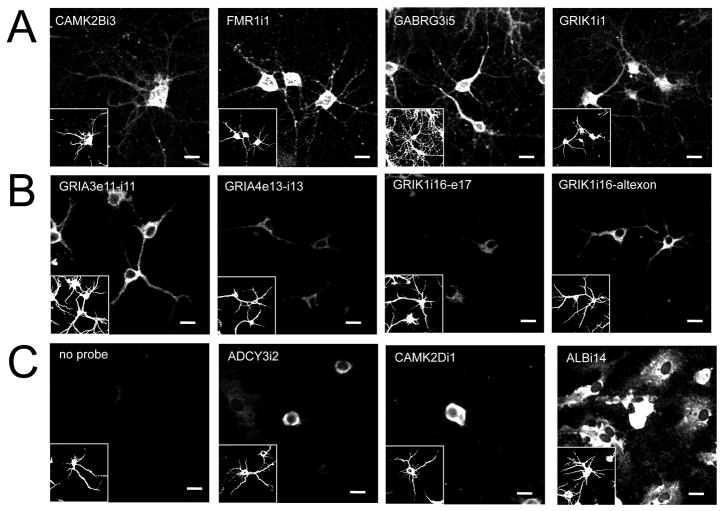
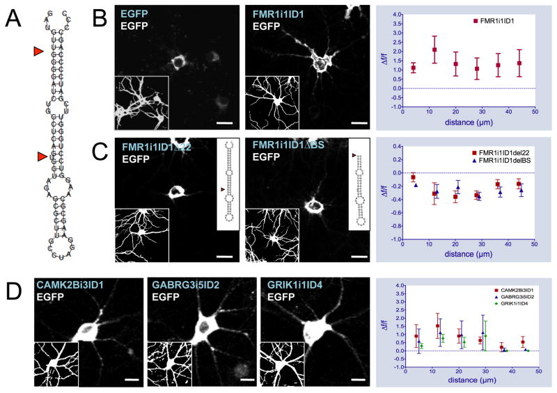
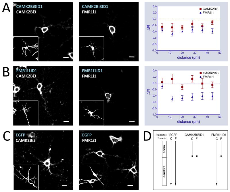
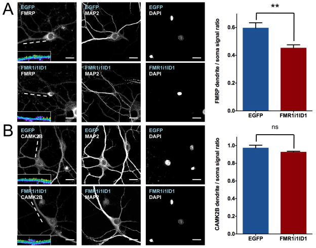
RNA precursors give rise to mRNA after splicing of intronic sequences traditionally thought to occur in the nucleus. Here, we show that intron sequences are retained in a number of dendritically-targeted mRNAs, by using microarray and Illumina sequencing of isolated dendritic mRNA as well as in situ hybridization. Many of the retained introns contain ID elements, a class of SINE retrotransposon. A portion of these SINEs confers dendritic targeting to exogenous and endogenous transcripts showing the necessity of ID-mediated mechanisms for the targeting of different transcripts to dendrites. ID elements are capable of selectively altering the distribution of endogenous proteins, providing a link between intronic SINEs and protein function. As such, the ID element represents a common dendritic targeting element found across multiple RNAs. Retention of intronic sequence is a more general phenomenon than previously thought and plays a functional role in the biology of the neuron, partly mediated by co-opted repetitive sequences.


Figures

Figure 1
Figure 1
In situ hybridization results for intronic probes on cultured rat hippocampal neurons. Panels are labeled according to intronic sequence detected using biotinylated (A) riboprobes or (B) oligo probes. Negative controls (C) for cells labeled in absence of probe (No probe) and with probes to non-retained introns are also included for reference (exon targeted and sense probe results found in Figure S2). Glia cells (MAP2 negative) are clearly positive for ALBi14 as shown. Riboprobe sequences are identical to those printed on intron microarrays (see Table S2). Oligo probes are 24-mers corresponding to genomic exon/intron splice junctions found in Illumina sequencing results for dendrites. Cells were also immunostained for MAP2 to identify neuronal soma and dendrites (insets). Scale bars = 20μm.
Figure 2
Figure 2
Intronic ID element sequences confer dendritic localization to reporter gene mRNA. Secondary structure of FMR1i1ID1 wild-type (A, inset for mutant sequences). In situ hybridizations with antisense biotinylated EGFP riboprobe on primary hippocampal neurons transfected with pEGFP-N1, FMR1i1ID1-EGFP wild-type (B), FMR1i1ID1 22-EGFP, and FMR1i1ID1 BS-EGFP mutant constructs (C). Blue text indicates transfected DNA construct, white text indicates in situ probe sequence. Insets represent MAP2 immunostaining (left) or secondary structure of FMR1i1ID1 mutants (right). Graphs at right represent in situ signal F/F (relative fluorescence signal difference) against distance from soma for FMR1i1ID1-EGFP wild-type versus pEGFP-N1 (top, expanded in Figure S3A–B), FMR1i1ID1 22-EGFP, and FMR1i1ID1 BS-EGFP mutants versus FMR1i1ID1-EGFP wild-type (bottom). (D) In situ hybridizations with antisense biotinylated EGFP riboprobe on hippocampal neurons transfected with CAMK2Bi3ID1ID1-EGFP, GABRG3i5ID2-EGFP, and GRIK1i1ID4-EGFP constructs. Graphs at right represent in situ signal F/F against distance from soma for each ID-EGFP construct versus pEGFP-N1. Scale bars = 20μm.
Figure 3
Figure 3
Intronic ID element sequences disrupt dendritic localization patterns of endogenous mRNA. In situ hybridizations with antisense biotinylated intron riboprobes on primary hippocampal neurons transfected with CAMK2Bi3ID1-EGFP (A), FMR1i1ID1-EGFP (B), pEGFP-N1 (C) constructs (GABRG3i5ID2-EGFP and GRIK1i1ID4 results found in Figure S4). Blue text indicates transfected DNA construct, white text indicates in situ probe sequence. Graphs at right represent in situ signal F/F against distance from soma for ID-EGFP constructs versus pEGFP-N1 using CAMK2Bi3 (left) and FMR1i1 (right) riboprobes. Insets represent MAP2 immunostaining. Scale bars = 20μm. (D) Schematic of ID cross competition results. Transfection labels indicate transfected DNA constructs, transcript labels indicate endogenous intron-retaining transcripts: C is CAMK2Bi3, F is FMR1i1. Arrows indicate endogenous intron-retaining transcript targeting from soma to dendrites.
Figure 4
Figure 4
Intronic ID element sequences modify subcellular distributions of endogenous proteins. Immunofluorescence with antibodies to FMRP (A) and CAMK2B (B) on primary hippocampal neurons transfected with pEGFP-N1 and FMR1i1ID1-EGFP constructs. Blue text indicates transfected DNA construct, white text indicates antibody target. Graphs at right represent immuno signal as ratios of dendrite to soma with SEM indicated. Mean dendritic to somal signal ratio +/− SEM for FMRP immunostaining: EGFP 0.56 +/− 0.04 (n=10), FMR1i1ID1 0.45 +/− 0.02 (n=13), t-test p = 0.0031. Mean dendritic to somal signal ratio +/− SEM for CAMK2B immunostaining: EGFP 0.97 +/− 0.03 (n=7), FMR1i1ID1 0.93 +/− 0.01 (n=9), t-test p = 0.16. Insets are three-dimensional topographical plots of signal intensity for the indicated dendrites (white lines) on a 0 (dark blue) to 2000 (yellow) pseudocolor range, showing a decreased signal for FMRP along the length of the FMR1i1ID1-transfected dendrite compared to the EGFP-transfected dendrite; no such difference is observed for CAMK2B. Scale bars = 20μm.

References

    1. Aakalu G, Smith WB, Nguyen N, Jiang C, Schuman EM. Dynamic visualization of local protein synthesis in hippocampal neurons. Neuron. 2001;30:489–502. - PubMed
    1. Altschul SF, Gish W, Miller W, Myers EW, Lipman DJ. Basic local alignment search tool. J Mol Biol. 1990;215:403–410. - PubMed
    1. Andreassi C, Riccio A. To localize or not to localize: mRNA fate is in 3′UTR ends. Trends Cell Biol. 2009;19:465–474. - PubMed
    1. Baskerville S, Bartel DP. Microarray profiling of microRNAs reveals frequent coexpression with neighboring miRNAs and host genes. RNA. 2005;11:241–247. - PMC - PubMed
    1. Bell TJ, Miyashiro KY, Sul JY, Buckley PT, Lee MT, McCullough R, Jochems J, Kim J, Cantor CR, Parsons TD, Eberwine JH. Intron retention facilitates splice variant diversity in calcium-activated big potassium channel populations. Proceedings of the National Academy of Sciences of the United States of America. 2010;107:21152–21157. - PMC - PubMed

Publication types