. 2011 Mar 10;69(5):885-92.
doi: 10.1016/j.neuron.2011.02.006.

Synaptic integration gradients in single cortical pyramidal cell dendrites

Affiliations

Synaptic integration gradients in single cortical pyramidal cell dendrites

Tiago Branco et al. Neuron. .

Abstract

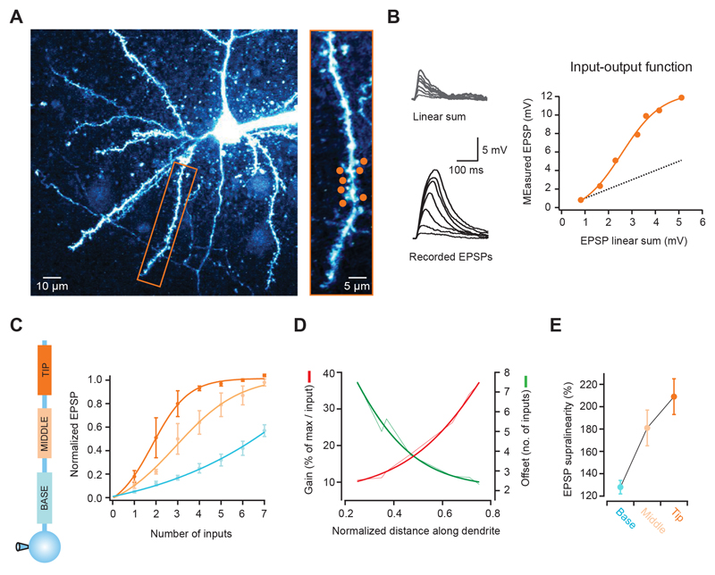
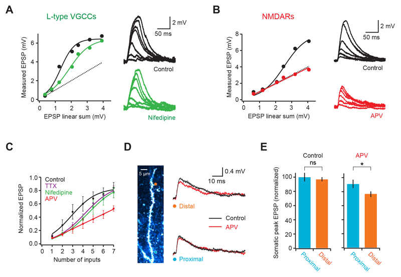
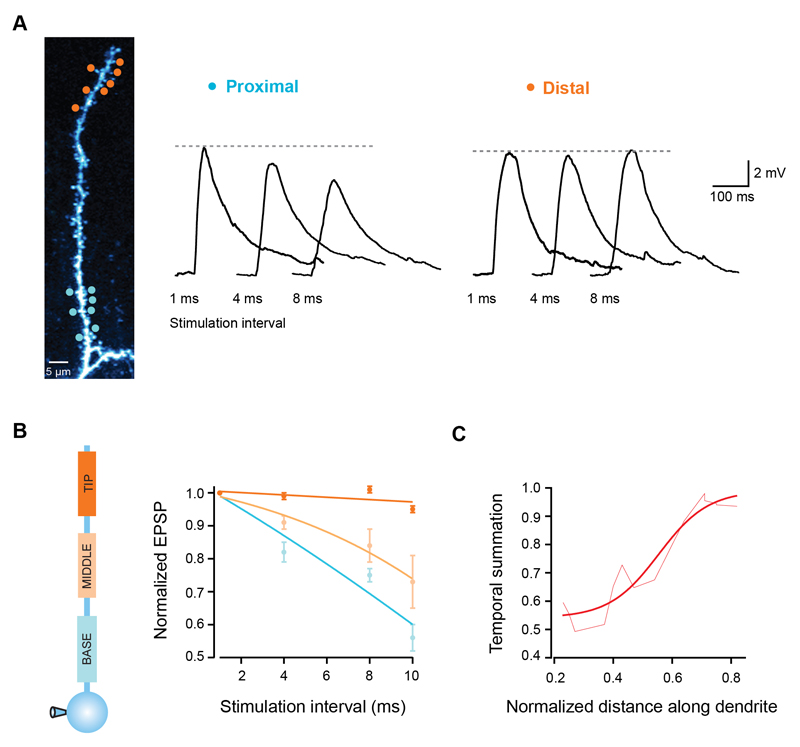
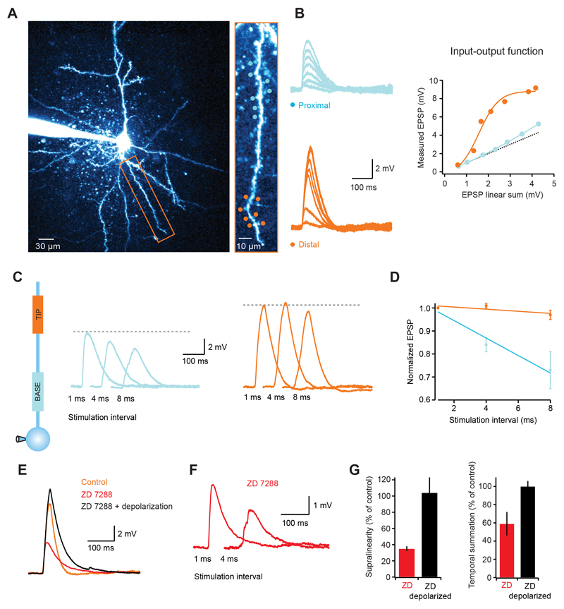
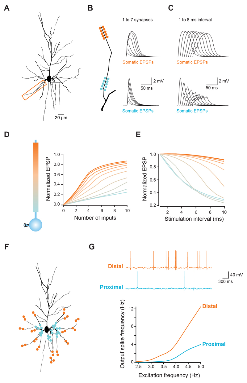
Cortical pyramidal neurons receive thousands of synaptic inputs arriving at different dendritic locations with varying degrees of temporal synchrony. It is not known if different locations along single cortical dendrites integrate excitatory inputs in different ways. Here we have used two-photon glutamate uncaging and compartmental modeling to reveal a gradient of nonlinear synaptic integration in basal and apical oblique dendrites of cortical pyramidal neurons. Excitatory inputs to the proximal dendrite sum linearly and require precise temporal coincidence for effective summation, whereas distal inputs are amplified with high gain and integrated over broader time windows. This allows distal inputs to overcome their electrotonic disadvantage, and become surprisingly more effective than proximal inputs at influencing action potential output. Thus, single dendritic branches can already exhibit nonuniform synaptic integration, with the computational strategy shifting from temporal coding to rate coding along the dendrite.


Figures

Figure 1
Figure 1. The input-output function varies with distance along single pyramidal cell dendrites
(A) Left, 2-photon image of a layer 2/3 neuron pyramidal filled with Alexa 594. Rectangular box indicates basal dendrite selected for experiment. Right, selected dendrite with 7 glutamate uncaging spots (orange circles). (B) Somatic voltage responses to increasing number of stimulated synapses (from dendrite and spots shown in a, activated at 1 ms intervals). Bottom traces show recorded responses, and top traces the linear sum expected from the individual responses to each spot. The graph on the right shows that the recorded peak EPSPs are markedly supralinear and grow as a sigmoid function (dotted line is the linear sum, orange circles the actual response; orange line is a fit to the data). (C) Differences in the input-output function according to the position along individual dendrites. Lines are sigmoid fits to the data, and values are shown normalized to the maximum of the fit. Distal synapses have a higher gain function, which is also shifted to the right (summarized in D). (E) EPSP supralinearity also increases with distance from the soma (values for activation of 3 inputs).
Figure 2
Figure 2. Biophysical mechanism of dendritic supralinear integration
(A) Somatic voltage response to increasing number of synapses on a basal dendrite before (black) and after blocking L-type calcium channels with Nifedipine (green). Circles are datapoints, thick lines are fits to the data and dotted line is the linear sum. (B) Similar to A but comparing somatic responses before (black) and after (red) blocking NMDA receptors with APV. Note that responses become linear. (C) Summary plot (pooled data from multiple cells, n = 9 for control) showing that both Nifedipine (n = 6) and TTX (n = 4) shift the dendritic input-output curve to right, while APV (n = 5) linearizes it. (D) Activation of either a distal (orange circle) or a proximal (blue circle) single spine produces a similar size EPSP at the soma (right, black traces). Block of NMDARs reveals a larger NMDA component in the distal EPSP (right, red traces). (E) Summary data comparing EPSP size and NMDA content between proximal and distal spines (n = 8).
Figure 3
Figure 3. Temporal summation gradient along pyramidal cell dendrites
(A) Seven uncaging spots were placed either at the tip (orange circles) or close to the branchpoint of a single dendrite (blue circles), and activated with different degrees of synchrony. Traces show somatic EPSPs in response to increasing stimulation intervals for both locations. Note the invariance of the EPSP peak for distal synapses. (B) EPSP peak normalized to the response for 1 ms intervals for 3 different regions of single dendrites. Lines are fits to the data. (C) Temporal summation increases towards the dendritic tip (measured as the EPSP peak at 10 ms interval normalized to the response at 1 ms interval). Smooth line is a sigmoid fit.
Figure 4
Figure 4. Dendritic integration gradients in layer 5 pyramidal neurons
(A) Left, 2-photon image of a layer 5 pyramidal cell with the dendrite targeted in experiment indicated by rectangular box. Right, targeted dendrite with distal (orange circles) and proximal (blue circles) uncaging spots. (B) Somatic EPSPs in response to increasing numbers of stimulated spines (1 ms interval) for the proximal (top, blue) and distal (bottom, orange) locations. The graph on the right shows that, like in layer 2/3 cells, distal synapses have a highly supralinear and sigmoidal input-output function, while proximal locations show a much more linear function. (C) Somatic voltage traces for stimulation at increasing intervals at proximal (blue traces) and distal (orange traces) locations. (D) Summary data showing that temporal summation is more effective at distal locations. (E) Blocking Ih channels decreases the EPSP amplitude (red trace), which is restored upon somatic depolarization (black trace). Note the difference in the EPSP decay between the black and orange (control) traces, illustrating the Ih-dependent speeding of EPSP decay. (F) Somatic EPSP for two stimulation intervals of distal synapses in Ih block. Note how the response at 4 ms is significantly smaller than at 1 ms (compare with orange traces in (C). (G) Summary data showing the effects of Ih block on dendritic supralinearity and efficiency of temporal summation.
Figure 5
Figure 5. Modelling the impact of dendritic integration gradients on neuronal output
(A) Morphology of a reconstructed layer 2/3 pyramidal used for simulations. Box indicates dendrite used in B-E. (B,C) Clusters of synapses were placed at different locations along the dendrite (total length = 90 μm). (B) Increasing number of synapses were activated or (C) all synapses activated at different intervals. Traces show somatic voltage responses for proximal (bottom) and distal synapses (top), which reproduce the experimental data (see Figure 1A and 2A). (D,E) Summary for all tested locations in the model, showing the same gradients for the gain of the input-output function (D) and temporal summation (E) that were observed experimentally. (F) Schematic illustration of 169 synapses randomly distributed across all basal dendrites, with either a distal or a proximal bias. (G) Each synapse was activated with independent Poisson trains of increasing frequency and the somatic voltage monitored. As the excitation frequency increases, the spiking frequency increases more rapidly for distally distributed synapses. Traces show responses for distal (orange) and proximal (blue) distributions stimulated at 4 Hz.

References

    1. Abeles M. Corticonics: neural circuits of the cerebral cortex. Cambridge, UK: Cambridge University Press; 1991.
    1. Andersen P, Morris R, Amaral D, Bliss TV, O'Keefe J. The Hippocampus Book. New York: Oxford University Press; 2006.
    1. Andersen P, Silfvenius H, Sundberg SH, Sveen O. A comparison of distal and proximal dendritic synapses on CA1 pyramids in guinea-pig hippocampal slices in vitro. The Journal of Physiology. 1980;307:273–299. - PMC - PubMed
    1. Berger T, Larkum ME, Lüscher HR. High I(h) channel density in the distal apical dendrite of layer V pyramidal cells increases bidirectional attenuation of EPSPs. J Neurophysiol. 2001;85:855–68. - PubMed
    1. Branco T, Häusser M. The single dendritic branch as a fundamental functional unit in the nervous system. Curr Opin Neurobiol. 2010;20:494–502. - PubMed

Publication types

LinkOut - more resources