. 2011 Mar 10;69(5):918-29.
doi: 10.1016/j.neuron.2011.01.030.

Phosphorylation regulates OLIG2 cofactor choice and the motor neuron-oligodendrocyte fate switch

Affiliations

Phosphorylation regulates OLIG2 cofactor choice and the motor neuron-oligodendrocyte fate switch

Huiliang Li et al. Neuron. .

Abstract

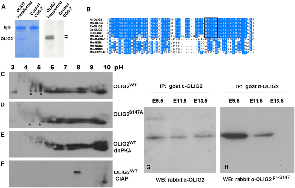
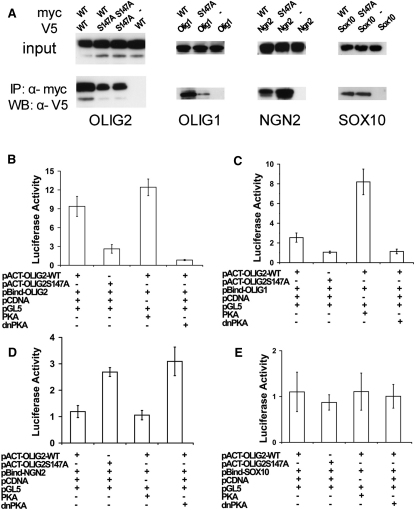
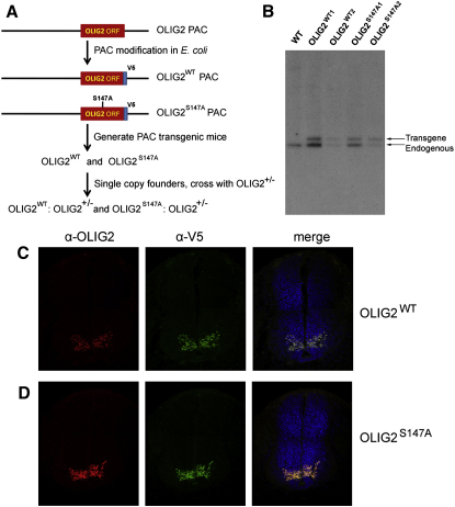
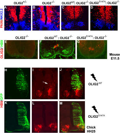
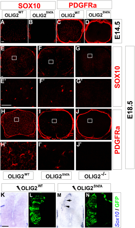
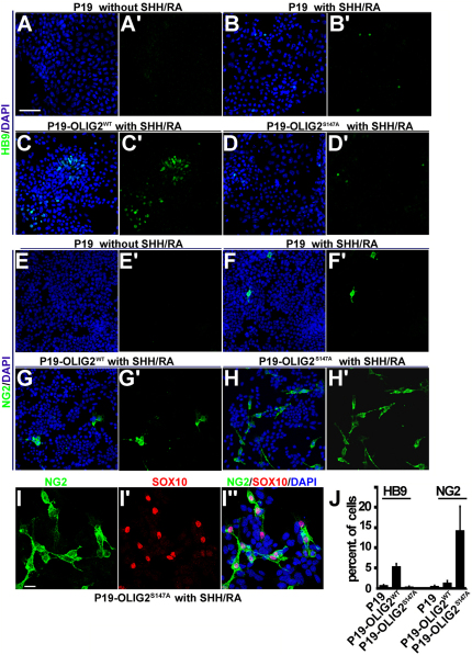
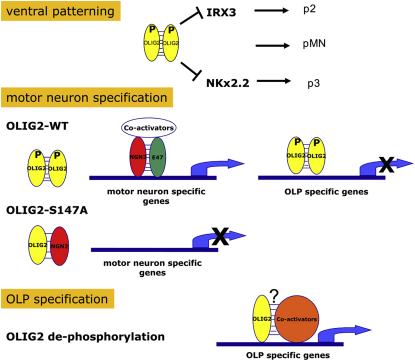
A fundamental feature of central nervous system development is that neurons are generated before glia. In the embryonic spinal cord, for example, a group of neuroepithelial stem cells (NSCs) generates motor neurons (MNs), before switching abruptly to oligodendrocyte precursors (OLPs). We asked how transcription factor OLIG2 participates in this MN-OLP fate switch. We found that Serine 147 in the helix-loop-helix domain of OLIG2 was phosphorylated during MN production and dephosphorylated at the onset of OLP genesis. Mutating Serine 147 to Alanine (S147A) abolished MN production without preventing OLP production in transgenic mice, chicks, or cultured P19 cells. We conclude that S147 phosphorylation, possibly by protein kinase A, is required for MN but not OLP genesis and propose that dephosphorylation triggers the MN-OLP switch. Wild-type OLIG2 forms stable homodimers, whereas mutant (unphosphorylated) OLIG2(S147A) prefers to form heterodimers with Neurogenin 2 or other bHLH partners, suggesting a molecular basis for the switch.


Figures

Figure 1
Figure 1
OLIG2 Is Phosphorylated on S147 (A) In vitro [33P] incorporation assay. Cell lysates of Olig2-transfected Cos-7 cells were immunoprecipitated with rabbit anti-Myc, separated by SDS-PAGE, and stained with Coomassie blue (left panel). The gel was dried and autoradiographed (right panel). OLIG2 bands are indicated (arrowheads). (B) Sequence alignment of bHLH domain of OLIG2 and its relatives. A predicted PKA phosphorylation motif containing S147 (black box) was found in the OLIG1/2 bHLH domain (helix 2) of different species. Hs, Homo sapiens; Mm, Mus musculus; Rn, Rattus norvegicus (brown rat); Gg, Gallus gallus (chicken); Dr, Danio rerio (zebrafish). (C–F) Transfected Cos-7 cells were analyzed by 2D PAGE followed by WB with anti-Myc antibody: (C) Myc-tagged OLIG2WT; (D) OLIG2S147A; (E) OLIG2WT with dnPKA; and (F) OLIG2WT lysate treated with CIAP. (G and H) OLIG2-S147 was phosphorylated in vivo. Mouse spinal cord homogenates from different developmental stages were analyzed by IP with goat anti-OLIG2, followed by WB with (G) rabbit anti-OLIG2 or (H) rabbit anti-OLIG2 ph-S147. See also Figure S1 and Table S1.
Figure 2
Figure 2
S147A Mutation Alters the Dimerization Properties of OLIG2 (A) Cos-7 cells were cotransfected with expression constructs encoding Myc-tagged OLIG2WT or OLIG2S147A together with V5-tagged OLIG2, OLIG1, NGN2, or SOX10. Cell lysates were immunoprecipitated with rabbit anti-Myc, followed by WB with anti-V5. In parallel, one-twentieth of each cell lysate was subjected directly to WB with anti-OLIG2. (B–E) Luciferase assay using cell lysates of transfected Cos-7 cells. The amounts of transfected DNA were normalized with pCDNA empty vector; Renilla Luciferase activity was used to calibrate transfection efficiency. Results are displayed as fold-increase of firefly Luciferase activity compared to controls (mean ± SEM of three independent experiments). See also Figure S2.
Figure 3
Figure 3
Construction and Characterization of OLIG2S147A Transgenic Mice (A) The OLIG2 open reading frame in a mouse Olig2 PAC was modified to contain a single mutation Serine147 → Alanine and a downstream V5-tag (Olig2S147A). A V5-tagged wild-type sequence was made as a control (Olig2WT). (B) Identification of single-copy transgenic founders by Southern blot. The transgene band was normalized to the endogenous Olig2 band; founders whose transgene band was less intense than the endogenous band carried one copy of the Olig2 transgene. (C and D) Transgenic embryos expressed OLIG2 faithfully in the ventral spinal cord. Transverse sections of E11.5 spinal cords from Olig2WT (C) and Olig2S147A (D) transgenic embryos were coimmunolabeled with OLIG2 (red) and the V5 epitope (green).
Figure 4
Figure 4
Ventral Patterning and MN Generation Are Disrupted in Olig2S147A Mice The pMN progenitor domain normally occupies the gap between PAX6 (red) and NKX2.2 (blue) immunolabeling in the ventral spinal cord of E11.5 embryos (A and C). pMN was missing in both Olig2 null and Olig2S147A mice (B and D). MNs were immunolabeled with anti-HB9 (green) in the Olig2WT spinal cord (F) but were absent in Olig2 null (E) and Olig2S147A (G). OLIG2S147A protein (red) was still expressed in Olig2S147A mice (G). (H–M) Olig2 expression constructs were electroporated in the HH12–14 chick neural tube. Electroporated cells were visualized 24 hr later by GFP immunolabeling (green). Ectopic HB9 expression (red) was induced by Olig2WT (H–J, white arrow), but not Olig2S147A (K–M). See also Figure S3.
Figure 5
Figure 5
OL Specification in Olig2S147A and Olig2WT Mice OL lineage cells were marked by SOX10 or PDGFRa immunolabeling (red). OLPs were present in Olig2WT mice at E14.5 (A and C), but not in Olig2S147A mice (B and D). At E18.5, SOX10- and PDGFRa-positive OLPs were detected in spinal cords of Olig2S147A mice (F, F′, I, and I′), though in reduced numbers compared to Olig2WT controls (E, E′, H, and H′). No OLPs whatsoever were detected in Olig2 null mice (G, G′, J, and J′). (E′–G′) and (H′–J′) are high-magnification views of the areas marked in (E)–(G) and (H)–(J), respectively. (K–N) Olig2 expression constructs together with a GFP expression vector were electroporated into HH12–14 chick neural tubes, and electroporated cells were visualized 48 hr later by GFP immunolabeling (green). Ectopic Sox10 expression (blue, arrows) was induced by transfected Olig2S147A (M and N), but not by Olig2WT (K and L). Scale bars, 80 μm (A–D); 50 μm (E–J′); 100 μm (K–N). See also Figure S4.
Figure 6
Figure 6
OLIG2S147A Promotes OL Fate Specification but Represses MN Fate in Cultured P19 Cells (A–D) MN induction: addition of SHHAg1.2 agonist and RA induced aggregated P19 cells to express the MN marker HB9 (B and B′). This induction was accentuated in stable cell line P19-OLIG2WT (C and C′) but repressed in P19-OLIG2S147A (D and D′). (E–H) OL induction: SHHAg1.2 and RA also induced aggregated P19 cells to express OLP marker NG2 (F and F′). This induction was slightly amplified in the P19-OLIG2WT line (G and G′) but dramatically magnified in P19-OLIG2S147A (H and H′). (I, I′, and I″) The NG2-positive cells (green) that were induced in P19-OLIG2S147A cultures also expressed SOX10 (red), confirming them as OL lineage cells. Cell nuclei were counterstained with DAPI (blue). (J) Quantification of HB9 and NG2-inducing activity. Scale bar, 50 μm (A–H′ and I–I″). See also Figure S5.
Figure 7
Figure 7
A Model of How OLIG2 Might Sequentially Regulate MN and OLP Fates through Reversible Phosphorylation on S147 A key feature is that dephosphorylation of OLIG2 increases the efficiency of OLIG2-NGN2 heterodimer formation, reducing the amount of NGN2 available for activating MN-specific genes and thereby contributing to the neuron-OL fate switch.

Comment in

References

    1. Arnett H.A., Fancy S.P., Alberta J.A., Zhao C., Plant S.R., Kaing S., Raine C.S., Rowitch D.H., Franklin R.J., Stiles C.D. bHLH transcription factor Olig1 is required to repair demyelinated lesions in the CNS. Science. 2004;306:2111–2115. - PubMed
    1. Bai C.B., Stephen D., Joyner A.L. All mouse ventral spinal cord patterning by hedgehog is Gli dependent and involves an activator function of Gli3. Dev. Cell. 2004;6:103–115. - PubMed
    1. Berg R.W., McBurney M.W. Cell density and cell cycle effects on retinoic acid-induced embryonal carcinoma cell differentiation. Dev. Biol. 1990;138:123–135. - PubMed
    1. Blom N., Gammeltoft S., Brunak S. Sequence and structure-based prediction of eukaryotic protein phosphorylation sites. J. Mol. Biol. 1999;294:1351–1362. - PubMed
    1. Blom N., Sicheritz-Ponten T., Gupta R., Gammeltoft S., Brunak S. Prediction of post-translational glycosylation and phosphorylation of proteins from the amino acid sequence. Proteomics. 2004;4:1633–1649. - PubMed

Publication types

MeSH terms

Substances