. 2011 Mar 22;123(11):1205-15.
doi: 10.1161/CIRCULATIONAHA.110.963314. Epub 2011 Mar 7.

Signal transducers and activators of transcription-3/pim1 axis plays a critical role in the pathogenesis of human pulmonary arterial hypertension

Affiliations

Signal transducers and activators of transcription-3/pim1 axis plays a critical role in the pathogenesis of human pulmonary arterial hypertension

Roxane Paulin et al. Circulation. .

Abstract

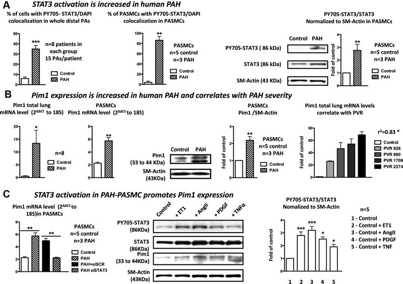
Background: Pulmonary artery hypertension (PAH) is a proliferative disorder associated with enhanced pulmonary artery smooth muscle cell proliferation and suppressed apoptosis. The sustainability of this phenotype required the activation of a prosurvival transcription factor like signal transducers and activators of transcription-3 (STAT3) and nuclear factor of activated T cell (NFAT). Because these factors are implicated in several physiological processes, their inhibition in PAH patients could be associated with detrimental effects. Therefore, a better understanding of the mechanism accounting for their expression/activation in PAH pulmonary artery smooth muscle cells is of great therapeutic interest.

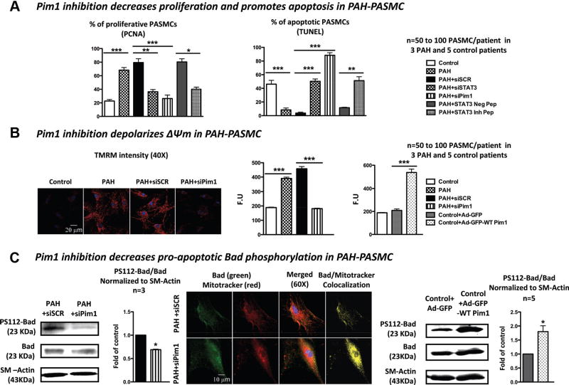
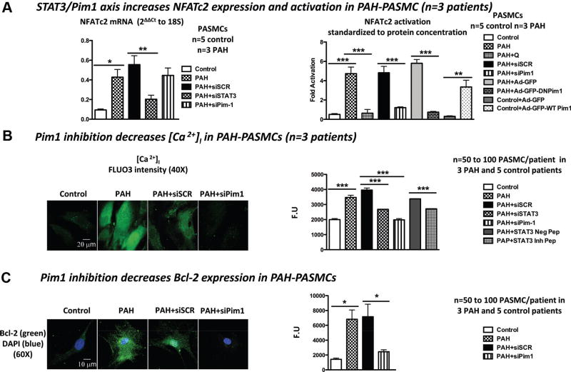
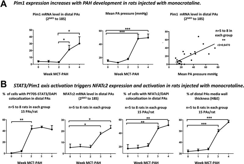
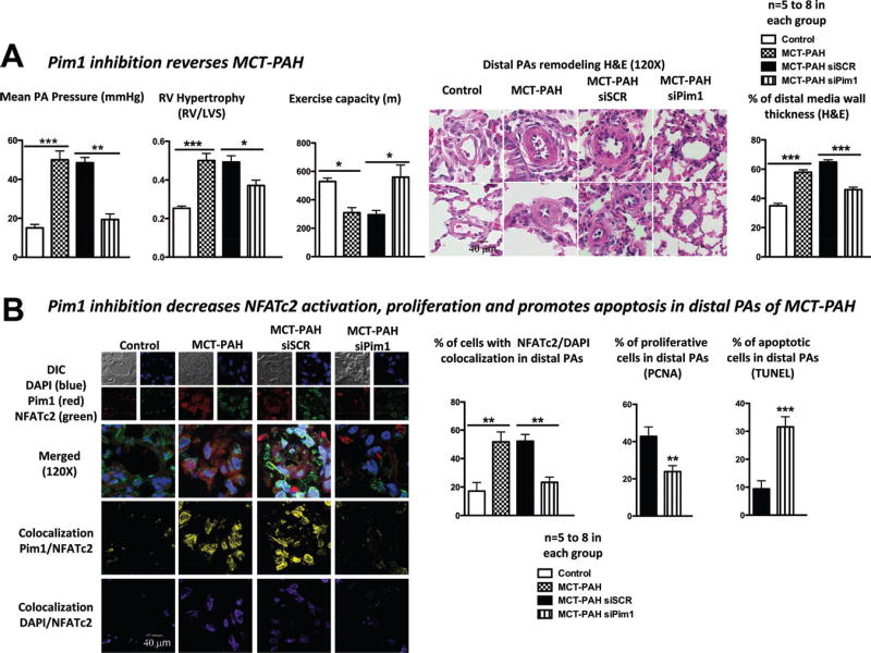
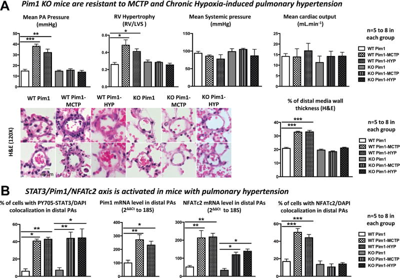
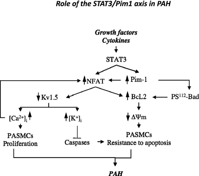
Methods and results: Using multidisciplinary and translational approaches, we demonstrated that STAT3 activation in both human and experimental models of PAH accounts for the expression of both NFATc2 and the oncoprotein kinase Pim1, which trigger NFATc2 activation. Because Pim1 expression correlates with the severity of PAH in humans and is confined to the PAH pulmonary artery smooth muscle cell, Pim1 was identified as an attractive therapeutic target for PAH. Indeed, specific Pim1 inhibition in vitro decreases pulmonary artery smooth muscle cell proliferation and promotes apoptosis, all of which are sustained by NFATc2 inhibition. In vivo, tissue-specific inhibition of Pim1 by nebulized siRNA reverses monocrotaline-induced PAH in rats, whereas Pim1 knockout mice are resistant to PAH development.

Conclusion: We demonstrated for the first time that inhibition of the inappropriate activation of STAT3/Pim1 axis is a novel, specific, and attractive therapeutic strategy to reverse PAH.


Figures

Figure 1
Figure 1
The signal transducers and activators of transcription-3 (STAT3)/Pim1 axis is activated in human pulmonary artery (PA) hypertension (PAH). A, STAT3 activation measured by PY705-STAT3 nuclear localization is significantly increased in both whole PAs and PA smooth muscle cells (SMCs) isolated from PAH patients compared with control. This was confirmed by immunoblots showing a 2.8-fold increase in the PY705-STAT3/STAT3 ratio in PAH PASMCs compared with control PASMCs. B, Pim1 mRNA levels measured by quantitative reverse-transcription polymerase chain reaction (qRT-PCR) are significantly increased in both lungs and PASMCs isolated from PAH patients compared with control patients. This was confirmed by immunoblots in isolated PASMCs. Finally, Pim1 mRNA levels in lungs correlate with PAH severity assessed by pulmonary vascular resistance (PVR). C, PAH PASMCs treated with siSTAT3 had a significant reduction in Pim1 expression (qRT-PCR) compared with the scrambled-treated cells. Endothelin-1– (ET1; 10 nmol/L), angiotensin II– (AngII; 200 nmol/L), platelet-derived growth factor– (PDGF; 30 ng/mL), and tumor necrosis factor– (TNF; 100 ng/mL) treated healthy PASMCs had an increased PY705-STAT3/STAT3 ratio associated with a proportional increase in Pim1 expression measured by immunoblots (P<0.05 vs control group).
Figure 2
Figure 2
Pim1 is implicated in the regulation of both pulmonary artery (PA) smooth muscle cell (PASMC) proliferation and apoptosis. A, PA hypertension (PAH) PASMCs had increased proliferation (percent proliferating cell nuclear antigen [PCNA]) and decreased apoptosis (percent terminal deoxynucleotidyl transferase–mediated dUTP nick-end labeling [TUNEL]) compared with healthy PASMCs. Both signal transducers and activators of transcription-3 inhibition and Pim1 inhibition significantly decrease PAH PASMC proliferation and promote apoptosis. B, Pim1 inhibition in PAH PASMCs reverses ΔΨm hyperpolarization (tetramethylrhodamine methyl-ester [TMRM]), whereas Pim1 activation in healthy PASMCs with adenovirus overexpressing Pim1 promotes it. C, Pim1 inhibition in PAH PASMCs decreases the PS112-Bad/Bad ratio, allowing Bad translocation to the mitochondria (increased colocalization between Bad in green and mitochondria [mitotracker red[ in red giving a yellow pattern).
Figure 3
Figure 3
The signal transducers and activators of transcription-3 (STAT3)/Pim1 axis accounts for nuclear factor of activated T cell-c2 (NFATc2) expression and activation in pulmonary artery hypertension (PAH) pulmonary artery smooth muscle cells (PASMCs). A, NFATc2 expression is increased in PAH PASMCs compared with control PASMCs. STAT3 inhibition decreases NFATc2 expression, whereas Pim1 inhibition has no effects. NFATc2 activation (luciferase assay) is increased in PAH PASMCs compared with control PASMCs. Pim1 inhibition decreases NFAT activation, whereas Pim1 overexpression in control PASMC promotes it. B, Pim1 inhibition in PAH PASMC decreases [Ca2+]i (FLUO3) and (C) Bcl-2 expression (immunofluorescence).
Figure 4
Figure 4
Pim1 expression is increased in the monocrotaline (MCT)-injected rat pulmonary artery (PA) hypertension (PAH) model. A, Heavy Pim1 mRNA expression is seen in PAs of rats with severe PAH (>21 days after MCT injection). In contrast, light expression was seen in mild PAH, and no expression was seen in rats with early PAH. Moreover, Pim1 expression correlates with PAH severity assed by mean PA pressure. B, Signal transducers and activators of transcription-3 (STAT3) activation (nuclear translocation of PY705-STAT3) precedes both Pim1 expression (quantitative reverse-transcription polymerase chain reaction [qRT-PCR]) and nuclear factor of activated T cell-c2 (NFATc2) expression (qRT-PCR) and activation (nuclear translocation), which paralleled both increased PA wall remodeling (hematoxylin and eosin staining) and PA pressure.
Figure 5
Figure 5
Pim1 inhibition reverses monocrotaline (MCT)–pulmonary artery (PA) hypertension (PAH). A, Pim1 inhibition (1 nebulization of 1 nmol) 18 days after MCT injection decreased mean PA pressure and right ventricular (RV) hypertrophy, improved exercise capacity, and diminished distal PA wall thickness. B, As in vitro, these effects were mediated by the inhibition of Pim1-dependent nuclear factor of activated T cell-c2 (NFATc2) activation showed by a decrease in Pim1/NFATc2 (yellow) and in NFATc2/DAPI (purple) colocalization, decreasing PA smooth muscle cell (PASMC) proliferation (percent proliferating cell nuclear antigen [PCNA]) and increasing apoptosis (percent terminal deoxynucleotidyl transferase–mediated dUTP nick-end labeling [TUNEL]).
Figure 6
Figure 6
Pim1 knockout (KO) mice are resistant to pyrrole-modified monocrotaline (MCTP) and chronic hypoxia (CH) pulmonary artery (PA) hypertension (PAH). A, Compared with Pim1 KO mice (n=5), MCTP and CH wild-type (WT) mice (n=8) developed PAH (increased PA pressure, right ventricular/left ventricle septum ratio, PA wall thickness) without detrimental effects on both systemic pressure and cardiac output. B, PAH development in MCTP WT and CH WT mice is due to the signal transducers and activators of transcription-3 (STAT3)/Pim1-dependent activation of nuclear factor of activated T cell-c2 (NFATc2). Both STAT3 activation (nuclear translocation assay) and NFATc2 mRNA levels (quantitative reverse-transcription polymerase chain reaction [qRT-PCR]) are increased in WT and KO MCTP and CH mice but, in the absence of Pim1, were not sufficient to trigger PAH.
Figure 7
Figure 7
Proposed mechanism of the role of the signal transducers and activators of transcription-3 (STAT3)/Pim1 axis in pulmonary artery (PA) hypertension (PAH). Schematic representation of STAT3/Pim1 implication in PAH pathogenesis. Increased circulating growth factors, agonists, and cytokines trigger STAT3 activation, resulting in increased nuclear factor of activated T cell-c2 (NFATc2) expression and increased Pim1 activation. Once activated, Pim1 triggers NFATc2 activation (nuclear translocation), promoting [Ca2+]i-dependent PA smooth muscle cell (PASMC) proliferation and inhibiting mitochondria-dependent apoptosis through a Bcl2-dependent mechanism. The NFATc2-dependent effect on Bcl2 will be reinforced by Pim1-dependent inhibition of Bad.

References

    1. Humbert M, Morrell NW, Archer SL, Stenmark KR, MacLean MR, Lang IM, Christman BW, Weir EK, Eickelberg O, Voelkel NF, Rabinovitch M. Cellular and molecular pathobiology of pulmonary arterial hypertension. J Am Coll Cardiol. 2004;43:13S–24S. - PubMed
    1. Archer S, Rich S. Primary pulmonary hypertension: a vascular biology and translational research “work in progress”. Circulation. 2000;102:2781–2791. - PubMed
    1. Ahmad S. Pulmonary hypertension and right heart failure. Chest. 1995;108:1773. - PubMed
    1. Bonnet S, Archer SL, Allalunis-Turner J, Haromy A, Beaulieu C, Thompson R, Lee CT, Lopaschuk GD, Puttagunta L, Bonnet S, Harry G, Hashimoto K, Porter CJ, Andrade MA, Thebaud B, Michelakis ED. A mitochondria-K+ channel axis is suppressed in cancer and its normalization promotes apoptosis and inhibits cancer growth. Cancer Cell. 2007;11:37–51. - PubMed
    1. Bonnet S, Rochefort G, Sutendra G, Archer SL, Haromy A, Webster L, Hashimoto K, Bonnet SN, Michelakis ED. The nuclear factor of activated T cells in pulmonary arterial hypertension can be therapeutically targeted. Proc Natl Acad Sci U S A. 2007;104:11418–11423. - PMC - PubMed

LinkOut - more resources