. 2011 May;79(5):1915-26.
doi: 10.1128/IAI.01270-10. Epub 2011 Mar 7.

Effect of cytokine interplay on macrophage polarization during chronic pulmonary infection with Cryptococcus neoformans

Affiliations

Effect of cytokine interplay on macrophage polarization during chronic pulmonary infection with Cryptococcus neoformans

Shikha Arora et al. Infect Immun. 2011 May.

Abstract

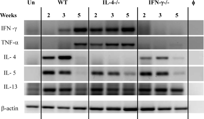
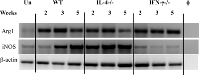
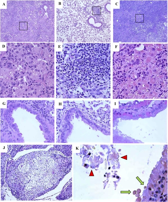
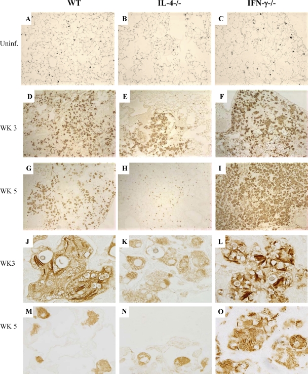
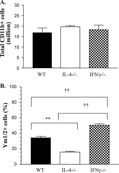
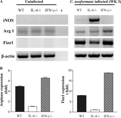
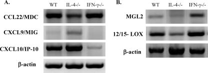
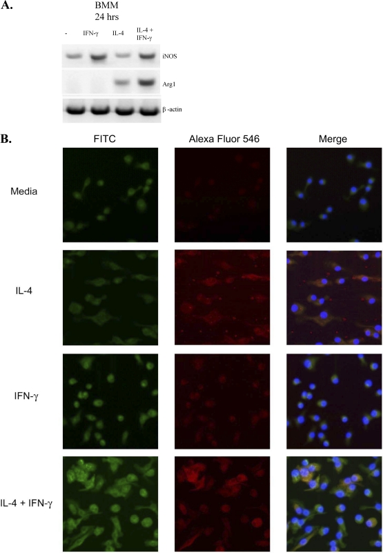
The immune response to Cryptococcus neoformans following pulmonary infection of C57BL/6 wild-type (WT) mice results in the development of persistent infection with characteristics of allergic bronchopulmonary mycosis (ABPM). To further clarify the role of Th1/Th2 polarizing cytokines in this model, we performed kinetic analysis of cytokine responses and compared cytokine profiles, pathologies, and macrophage (Mac) polarization status in C. neoformans-infected WT, interleukin-4-deficient (IL-4(-/-)), and gamma interferon-deficient (IFN-γ(-/-)) C57BL/6 mice. Results show that cytokine expression in the infected WT mice is not permanently Th2 biased but changes dynamically over time. Using multiple Mac activation markers, we further demonstrate that IL-4 and IFN-γ regulate the polarization state of Macs in this model. A higher IL-4/IFN-γ ratio leads to the development of alternatively activated Macs (aaMacs), whereas a higher IFN-γ/IL-4 ratio leads to the generation of classically activated Macs (caMacs). WT mice that coexpress IL-4 and IFN-γ during fungal infection concurrently display both types of Mac polarization markers. Concurrent stimulation of Macs with IFN-γ and IL-4 results in an upregulation of both sets of markers within the same cells, i.e., formation of an intermediate aaMac/caMac phenotype. These cells express both inducible nitric oxide synthase (important for clearance) and arginase (associated with chronic/progressive infection). Together, our data demonstrate that the interplay between Th1 and Th2 cytokines supports chronic infection, chronic inflammation, and the development of ABPM pathology in C. neoformans-infected lungs. This cytokine interplay modulates Mac differentiation, including generation of an intermediate caMac/aaMac phenotype, which in turn may support chronic "steady-state" fungal infection and the resultant ABPM pathology.


Figures

Fig. 1.
Fig. 1.
Effect of IL-4 and IFN-γ deletion on cytokine expression in C. neoformans-infected lungs. Total lung RNA was isolated from uninfected and C. neoformans-infected mice at 2, 3, and 5 wpi. RT PCR was performed for selected cytokines, and electrophoresis of PCR products was performed in a 2% agarose gel. Photographs show bands of the predicted sizes. φ, no-RNA control; Un, uninfected WT mice. Each lane is representative of 3 to 5 mice/group/time point. Baseline expression levels of cytokines were similar in uninfected lungs from WT, IL-4−/−, and IFN-γ−/− mice.
Fig. 2.
Fig. 2.
Effect of IL-4 and IFN-γ deletion on the kinetics of iNOS and arginase mRNA expression during C. neoformans infection. RNA from WT, IL-4−/−, and IFN-γ−/− mice was isolated and analyzed as described in the legend for Fig. 1. Photographs show bands of the predicted sizes for each PCR product. φ, no-RNA control; Un, uninfected WT mice. Each lane is representative of 3 to 5 mice/group/time point.
Fig. 3.
Fig. 3.
Effect of IL-4 and IFN-γ deletion on ABPM pathology in C. neoformans-infected lungs. Photomicrographs are H&E-stained sections of C. neoformans-infected lungs from WT (A, D, and G), IL-4−/− (B, E, and H), and IFN-γ−/− (C, F, and I to K) mice at 5 wpi. Low-power images (A, B, and C) demonstrate differences in the extent of inflammatory responses and consolidation of lung tissue (10× objective). High-power images (D, E, and F) of the boxed areas demonstrate cellular composition and differences in Mac morphology (40× objective). Note the absence of microbe in IL-4−/− lungs and the increased microbial presence and accumulation of eosinophilic deposits in the Mac in IFN-γ−/− lungs. High-power images of airway sections (G, H, and I) demonstrate differences in airway epithelium. Note the presence of eosinophilic inclusions in the epithelial cells of IFN-γ−/− mice (I). Severe pulmonary pathology in IFN-γ−/− lungs is highlighted by the formation of cryptococcomas (J, 10× objective), the presence of eosinophilic crystals in the airway lumen (K, 40× objective, red arrowheads), and the secretion of eosinophilic inclusions in the airway lumen, or hyalinosis (K, green arrows).
Fig. 4.
Fig. 4.
Effects of IFN-γ and IL-4 deletion on regulation of Ym1/2 production by Mac in uninfected and C. neoformans-infected lungs. Sections of uninfected WT, IL-4−/−, and IFN-γ−/− mouse lungs (A, B, and C) and C. neoformans-infected lungs (D to O) were immunostained with anti-Ym1/2 antibody and developed with a peroxidase-linked detection system. Sections shown in panels A to F and J to L were photographed using a 10× objective, while those in panels G to I and M to O were photographed using a 100× objective. Note that the basal level of Ym1/2 production by Mac in the uninfected lungs was not affected by IL-4 or IFN-γ deletion; however, accumulation of Ym1/2-positive Mac and intracellular Ym1/2 crystal deposition are ablated in IL-4−/− and augmented in IFN-γ−/− infected lungs.
Fig. 5.
Fig. 5.
Effects of IFN-γ and IL-4 deletion on numbers of Ym1/2-expressing CD11b+ cells in C. neoformans-infected lungs. (A) CD11b-expressing cells were isolated from C. neoformans-infected WT, IL-4−/−, and IFN-γ−/− mice (3 wpi) by MACS separation and counted under a microscope. The data are means from 3 independent experiments, with ≥3 mice/group/experiment. (B) CD11b-selected cells were stained with Ym1/2 Ab to enumerate Ym1/2-positive and -negative cells. Approximately 100 cells were counted in 10 different fields on a slide. **, P < 0.005 for the IL-4−/− group compared to the WT group; ††, P < 0.005 for the IL-4−/− group compared to the IFN-γ−/− group. Note that the total number of CD11b-selected cells was not affected by IL-4 or IFN-γ deletion but that there was a strong effect on the numbers of cell staining positive for Ym1/2.
Fig. 6.
Fig. 6.
Effects of IFN-γ and IL-4 deletion on the expression of selected caMac and aaMac genes by Mac from uninfected and C. neoformans-infected mice. RNA from enriched Mac isolates was analyzed at 3 wpi. (A) RT PCR mRNA analysis of RNA from iNOS, Arg1, and Fizz1 was performed as described in the legend for Fig. 1. The band photographs are representative of 3 independent experiments. (B) RNA from each sample was subjected to semiquantitative RT PCR analysis for Arg1 and Fizz1, and expression was normalized to GAPDH (glyceraldehyde-3-phosphate dehydrogenase) housekeeping gene expression within each sample. The average relative expression of each gene in IL-4−/− cells in each independent experiment was used to calculate the relative expression ratios for WT and IFN-γ−/− cells. The graph is representative of 3 or more experiments, where each sample was run in triplicate (n = 3 mice/group).
Fig. 7.
Fig. 7.
Effects of IFN-γ and IL-4 deletion on the expression of aaMac- and caMac-specific markers by Mac from C. neoformans-infected mice. CD11b-selected cell isolates were analyzed for mRNA expression of chemokines (A) or MGL2 and 12/15-LOX (B) at 3 wpi. Photographs show the bands of predicted sizes and are representative of 3 or more independent experiments.
Fig. 8.
Fig. 8.
Effects of IFN-γ and IL-4 on induction of iNOS2 and Arg1 in BMM. BMM from WT mice were precultured and isolated as described in Materials and Methods and transferred to 8-well glass Labtek tissue culture plates at a cell density of 1 × 10⁵ cells/well for 24 h at 37°C and 5% CO2 in media alone or with recombinant mouse IFN-γ, IL-4, or a combination of both cytokines. (A) RNA from these cells was collected and analyzed by RT PCR. (B) Cells were fixed, permeabilized, incubated sequentially with anti-iNOS FITC-conjugated antibodies, anti-Arg1 Alexa Fluor 546 phalloidin (Imgenex, San Diego, CA), and DAPI (to stain cell nuclei), and analyzed with a Zeiss LSM510 confocal microscope. Note the red Arg1 staining in IL-4-treated BMM, the green iNOS staining in IFN-γ-treated BMM, and the ARG1/iNOS double-positive staining in BMM treated with a combination of IL-4/IFN-γ. The individual green- and red-channel pictures are presented in the left and middle columns, respectively; the merged images are presented in the right column.

References

    1. Alspaugh J. A., Granger D. L. 1991. Inhibition of Cryptococcus neoformans replication by nitrogen oxides supports the role of these molecules as effectors of macrophage-mediated cytostasis. Infect. Immun. 59:2291–2296 - PMC - PubMed
    1. Arora S., et al. 2005. Role of IFN-gamma in regulating T2 immunity and the development of alternatively activated macrophages during allergic bronchopulmonary mycosis. J. Immunol. 174:6346–6356 - PubMed
    1. Beamer G. L., et al. 2008. Interleukin-10 promotes Mycobacterium tuberculosis disease progression in CBA/J mice. J. Immunol. 181:5545–5550 - PMC - PubMed
    1. Bhan U., et al. 2008. Toll-like receptor 9 regulates the lung macrophage phenotype and host immunity in murine pneumonia caused by Legionella pneumophila. Infect. Immun. 76:2895–2904 - PMC - PubMed
    1. Brys L., et al. 2005. Reactive oxygen species and 12/15-lipoxygenase contribute to the antiproliferative capacity of alternatively activated myeloid cells elicited during helminth infection. J. Immunol. 174:6095–6104 - PubMed

Publication types

LinkOut - more resources