. 2011 May;79(5):1984-97.
doi: 10.1128/IAI.01077-10. Epub 2011 Mar 7.

Legionella pneumophila type II secretion dampens the cytokine response of infected macrophages and epithelia

Affiliations

Legionella pneumophila type II secretion dampens the cytokine response of infected macrophages and epithelia

Kessler McCoy-Simandle et al. Infect Immun. 2011 May.

Abstract

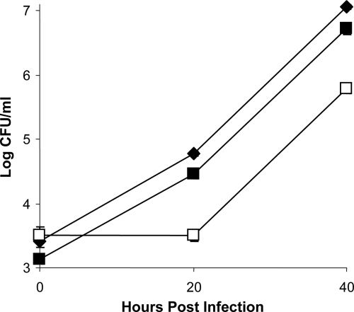
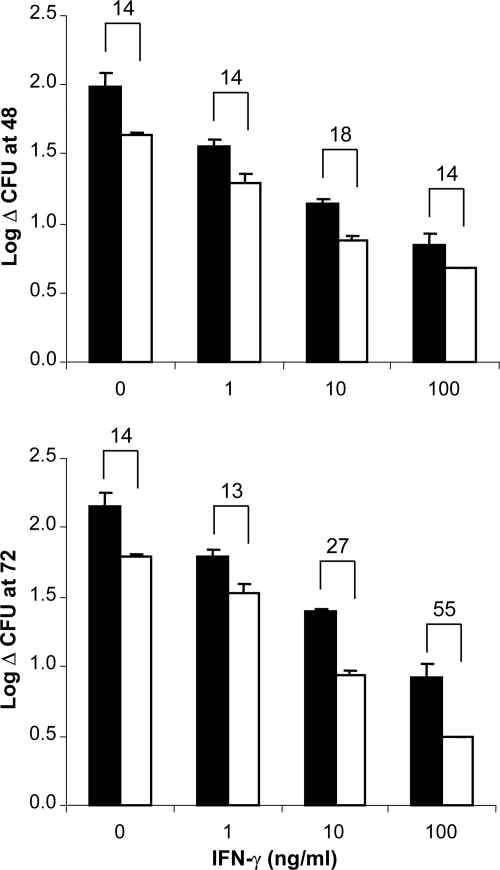
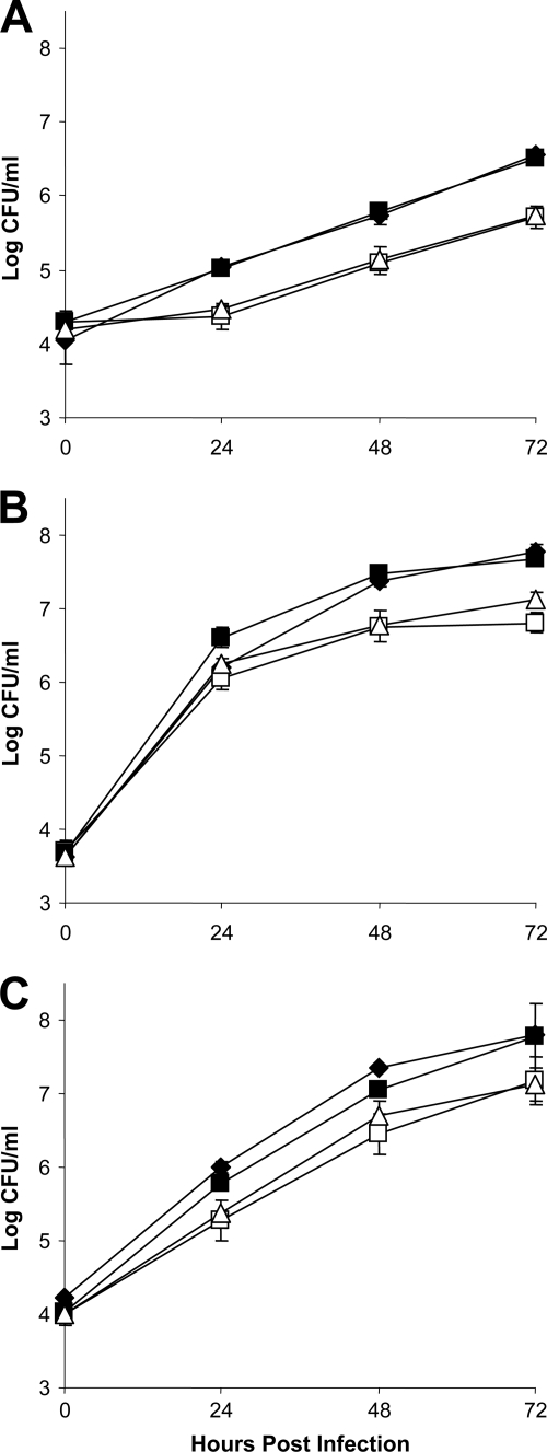
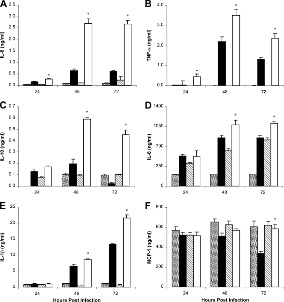
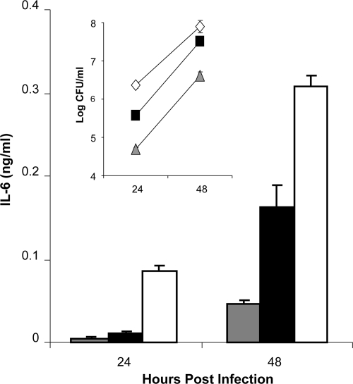
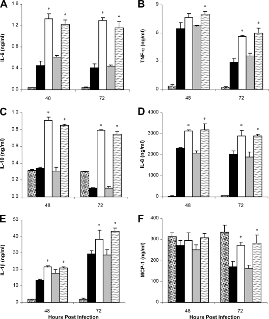
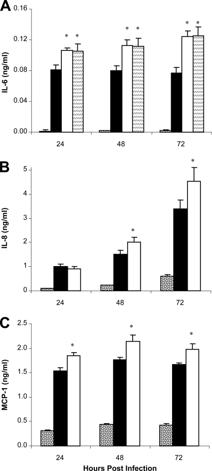
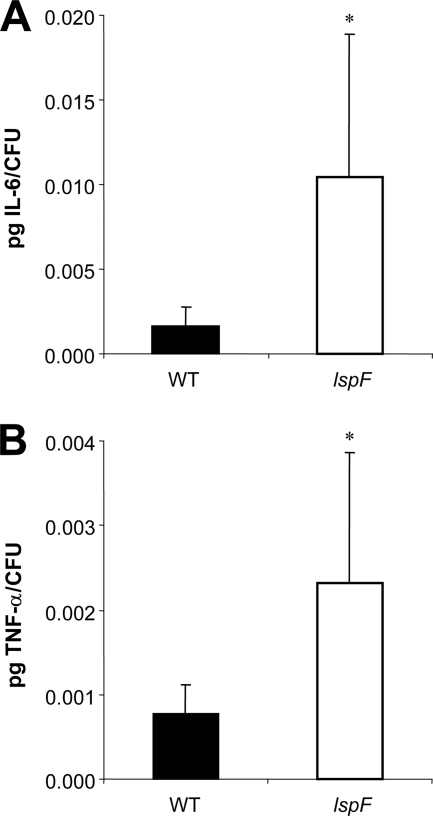
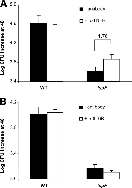
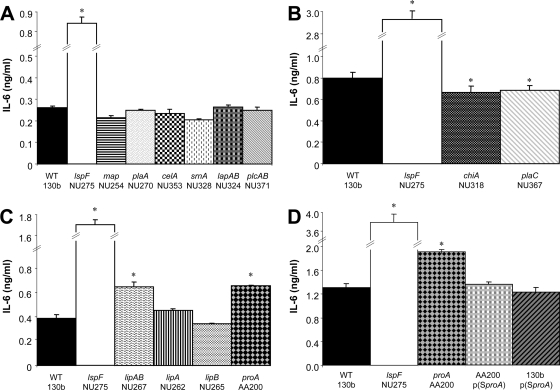
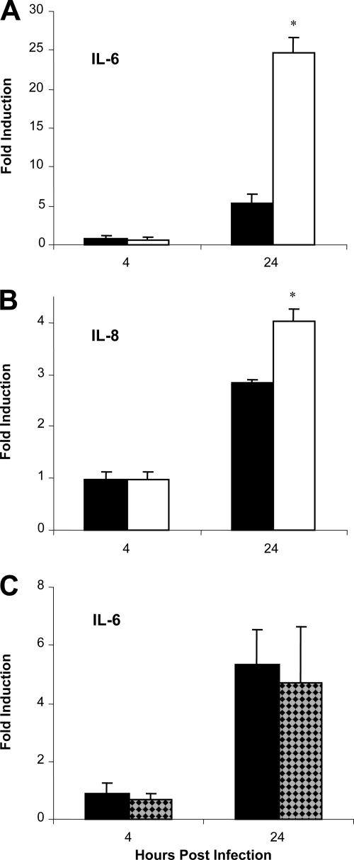
The type II secretion (T2S) system of Legionella pneumophila is required for the ability of the bacterium to grow within the lungs of A/J mice. By utilizing mutants lacking T2S (lsp), we now document that T2S promotes the intracellular infection of both multiple types of macrophages and lung epithelia. Following infection of macrophages, lsp mutants (but not a complemented mutant) elicited significantly higher levels of interleukin 6 (IL-6), tumor necrosis factor alpha (TNF-α), IL-10, IL-8, IL-1β, and MCP-1 within tissue culture supernatants. A similar result was obtained with infected lung epithelial cell lines and the lungs of infected A/J mice. Infection with a mutant specifically lacking the T2S-dependent ProA protease (but not a complemented proA mutant) resulted in partial elevation of cytokine levels. These data demonstrate that the T2S system of L. pneumophila dampens the cytokine/chemokine output of infected host cells. Upon quantitative reverse transcription (RT)-PCR analysis of infected host cells, an lspF mutant, but not the proA mutant, produced significantly higher levels of cytokine transcripts, implying that some T2S-dependent effectors dampen signal transduction and transcription but that others, such as ProA, act at a posttranscriptional step in cytokine expression. In summary, the impact of T2S on lung infection is a combination of at least three factors: the promotion of growth in macrophages, the facilitation of growth in epithelia, and the dampening of the chemokine and cytokine output from infected host cells. To our knowledge, these data are the first to identify a link between a T2S system and the modulation of immune factors following intracellular infection.


Figures

Fig. 1.
Fig. 1.
Intracellular infection of MH-S cell macrophages by wild-type and T2S mutant L. pneumophila. MH-S cells were inoculated with wild-type strain 130b (black diamonds), lspF mutant NU275 (white squares), or complemented mutant NU275 (pMF) (black squares), and then the number of CFU in each well was determined at various times postinfection. Results are the means and standard deviations of results from triplicate wells and are representative of three independent experiments. The differences in CFU recovery between the mutant and the wild type or the complemented mutant were significant at 20 and 40 h postinfection (Student's t test, P < 0.05).
Fig. 2.
Fig. 2.
Intracellular infection of BMD macrophages by wild-type and T2S mutant L. pneumophila. A/J mouse BMD macrophages that had been treated or not treated with the indicated concentrations of IFN-γ were inoculated with wild-type strain 130b (black bars) or lspF mutant NU275 (white bars), and then the number of CFU in each well was determined at various times postinfection. Bacterial growth is graphed as the increase in CFU number (in log units) from the time of inoculation to 48 h (top graph) or 72 h (bottom graph) postinoculation. Data are the means and standard deviations of results from triplicate wells and are representative of at least three independent experiments. The fold differences in CFU recovery between 130b- and NU275-infected monolayers, as indicated by the numbers over the brackets, were significant at both 48 and 72 h postinfection (Student's t test, P < 0.05). However, the magnitude of the differences between the strains did not change significantly as a result of the IFN-γ treatments (P > 0.05).
Fig. 3.
Fig. 3.
Intracellular infection of lung epithelial cells by wild-type and T2S mutant L. pneumophila. The A549 human alveolar type II epithelial cell line (A), WI-26 VA4 human alveolar type I epithelial cell line (B), and TC-1 murine alveolar epithelial cell line (C) were inoculated with wild-type strain 130b (black diamonds), lspF mutant NU275 (white squares), lspDE mutant NU258 (white triangles), or complemented mutant NU275 (pMF) (black squares), and then the number of CFU in each well was determined at various times postinfection. Results are the means and standard deviations of results from quadruplicate wells and are representative of three independent experiments. The differences in CFU recovery between the mutant and wild type or the complemented mutant were significant at 24, 48, and 72 h postinfection (Student's t test, P < 0.01).
Fig. 4.
Fig. 4.
Cytokine/chemokine output from macrophages infected with wild-type, T4S mutant, and T2S mutant L. pneumophila. U937 cells were either not infected (dotted bars) or infected (MOI = 0.5) with wild-type strain 130b (black bars), dotA mutant GG105 (diagonally striped bars), or lspF mutant NU275 (white bars), and then at the indicated times, the levels of IL-6 (A), TNF-α (B), IL-10 (C), IL-8 (D), IL-1β (E), and MCP-1 (F) in culture supernatants were determined by ELISA. Data are the means and standard deviations of results from triplicate wells and are representative of at least three independent experiments. The absence of some data bars in panel B is simply a reflection of the exceedingly low levels of TNF-α that were detected in some of the monolayers. Asterisks indicate those time points when the cytokine/chemokine levels elicited by the lspF mutant were significantly (Student's t test, P < 0.05) above those produced by wild-type infection.
Fig. 5.
Fig. 5.
Cytokine output from U937 cells infected with differing doses of wild-type L. pneumophila. U937 cells were infected with wild-type strain 130b at an MOI equal to 0.05 (gray triangles or bar), 0.5 (black squares or bar), or 5.0 (white diamonds or bar), and then at 24 and 48 h postinoculation, the numbers of CFU (inset) and amounts of secreted IL-6 within the infected wells were determined. Data are the means and standard deviations of results from triplicate wells and are representative of at least two independent experiments. At 24 and 48 h, IL-6 levels triggered by infections begun with an MOI of 5.0 were significantly greater than those produced by infections with the two lower doses of bacteria (Student's t test, P < 0.05). An MOI of 0.5 in turn elicited higher levels of the cytokine at 48 h than did an MOI of 0.05 (Student's t test, P < 0.05).
Fig. 6.
Fig. 6.
Cytokine/chemokine output from macrophages infected with T2S mutant and complemented T2S mutant L. pneumophila. U937 cells were either not infected (dotted bars) or infected (MOI = 0.5) with wild-type strain 130b (black bars), the lspF mutant NU275 (white bars), the complemented lspF mutant NU275(pMF) (gray bars), or the lspDE mutant NU258 (striped bars), and then at 48 and 72 h postinoculation the levels of IL-6 (A), TNF-α (B), IL-10 (C), IL-8 (D), IL-1β (E), and MCP-1 (F) in culture supernatants were determined by ELISA. Data are the means and standard deviations of results from triplicate wells and are representative of at least three independent experiments. Asterisks indicate those time points when the cytokine/chemokine levels elicited by the lspF and lspDE mutants were significantly (Student's t test, P < 0.05) above those produced by wild-type infection.
Fig. 7.
Fig. 7.
Cytokine/chemokine output from lung epithelial cells infected with wild-type and T2S mutant L. pneumophila. A549 cells were either not infected (dotted bars) or infected (MOI = 10) with wild-type strain 130b (black bars), lspF mutant NU275 (white bars), or lspDE mutant NU258 (striped bars [in panel A only]), and then at the indicated times the levels of IL-6 (A), IL-8 (B), and MCP-1 (C) in culture supernatants were determined by ELISA. Data are the means and standard deviations of results from triplicate wells and are representative of at least three independent experiments. Asterisks indicate points when the cytokine and chemokine levels elicited by the lspF and lspDE mutants were significantly (Student's t test, P < 0.05) above those produced by wild-type infection.
Fig. 8.
Fig. 8.
IL-6 and TNF-α levels in lungs infected with wild-type (WT) and T2S mutant L. pneumophila. A/J mice were intratracheally inoculated with equal numbers of either wild-type 130b (black bars) or the lspF mutant NU275 (white bars), and then at 24 h postinoculation, the levels of IL-6 and TNF-α per CFU in lung homogenates were determined by ELISA. Data are the means and standard deviations obtained for 9 to 10 infected animals and are representative of at least three independent experiments. The differences in cytokine levels between the wild-type- and mutant-infected lungs were significantly different (Student's t test, P < 0.05).
Fig. 9.
Fig. 9.
Effect of anticytokine antibodies on L. pneumophila growth in macrophages. Strain 130b (WT) and mutant NU275 (lspF) were used to infect U937 cells that had been treated in standard fashion (black bars) or exposed to antibodies (white bars) directed against either the TNF-α receptor (α-TNFR) (A) or the IL-6 receptor (B), and then at 0 and 48 h, the numbers of CFU in the monolayers were determined. Bacterial growth is graphed as the increase in numbers of CFU (in log units) from the time of inoculation to 48 h postinoculation. Data are the means and standard deviations of results from triplicate wells and are representative of at least three independent experiments. (A) The fold difference in mutant recovery between the untreated and treated monolayers indicated by the number over the bracket was significant (Student's t test, P < 0.05).
Fig. 10.
Fig. 10.
IL-6 output from macrophages infected with mutants of L. pneumophila lacking individual T2S-dependent effectors. (A to D) U937 cells were infected (MOI = 0.5) with wild-type strain 130b, the lspF mutant NU275, or mutants specifically lacking the indicated T2S substrate(s), and then at 72 h postinoculation, the levels of IL-6 in culture supernatants were determined by ELISA. Data are the means and standard deviations of results from triplicate wells and are representative of at least three independent experiments. Asterisks indicate those mutants that elicited a cytokine level that was significantly different (Student's t test, P < 0.05) from that triggered by the wild type. Related to the data in panel A, mutants NU268, NU369, and NU370, lacking just plcA or plcB, behaved similarly to plcA plcB double mutants, including strain NU371, which is shown in the figure, and did not show a phenotype that was distinct from that of the wild type (data not shown).
Fig. 11.
Fig. 11.
Cytokine transcripts from macrophages infected with wild-type and T2S mutant L. pneumophila. U937 cells were either not infected or infected with wild-type 130b (black bars in panels A to C), lspF mutant NU275 (white bars in panels A to B), or proA mutant AA200 (checkered bars in panel C), and then at 4 and 24 h postinoculation, the levels of IL-6 (A and C) and IL-8 (B) transcripts were determined by quantitative RT-PCR. Plotted on the y axis is the fold induction of the cytokine transcript in infected cells over the level of induction in the uninfected control. Data are means and standard deviations of results from triplicate wells and are representative of at least three independent experiments. Asterisks indicate statistically significant differences (Student's t test, P < 0.05) between the wild type and lspF mutant.

Similar articles

Cited by

References

    1. Akamine M., et al. 2005. Differential roles of Toll-like receptors 2 and 4 in in vitro responses of macrophages to Legionella pneumophila. Infect. Immun. 73:352–361 - PMC - PubMed
    1. Allard K. A., et al. 2009. Purification of legiobactin and the importance of this siderophore in lung infection by Legionella pneumophila. Infect. Immun. 77:2887–2895 - PMC - PubMed
    1. Amer A., et al. 2006. Regulation of Legionella phagosome maturation and infection through flagellin and host Ipaf. J. Biol. Chem. 281:35217–35223 - PubMed
    1. Amer A. O. 2010. Modulation of caspases and their non-apoptotic functions by Legionella pneumophila. Cell. Microbiol. 12:140–147 - PubMed
    1. Aragon V., Kurtz S., Cianciotto N. P. 2001. Legionella pneumophila major acid phosphatase and its role in intracellular infection. Infect. Immun. 69:177–185 - PMC - PubMed

Publication types

MeSH terms