. 2011 Apr 12;108(15):6294-9.
doi: 10.1073/pnas.1011866108. Epub 2011 Mar 7.

Universal rule for the symmetric division of plant cells

Affiliations

Universal rule for the symmetric division of plant cells

Sébastien Besson et al. Proc Natl Acad Sci U S A. .

Abstract

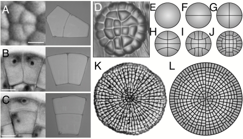
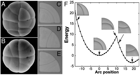
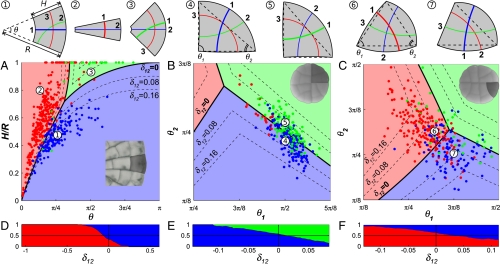
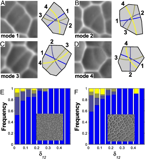
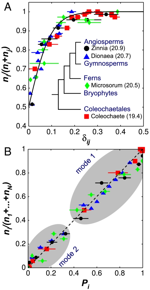
The division of eukaryotic cells involves the assembly of complex cytoskeletal structures to exert the forces required for chromosome segregation and cytokinesis. In plants, empirical evidence suggests that tensional forces within the cytoskeleton cause cells to divide along the plane that minimizes the surface area of the cell plate (Errera's rule) while creating daughter cells of equal size. However, exceptions to Errera's rule cast doubt on whether a broadly applicable rule can be formulated for plant cell division. Here, we show that the selection of the plane of division involves a competition between alternative configurations whose geometries represent local area minima. We find that the probability of observing a particular division configuration increases inversely with its relative area according to an exponential probability distribution known as the Gibbs measure. Moreover, a comparison across land plants and their most recent algal ancestors confirms that the probability distribution is widely conserved and independent of cell shape and size. Using a maximum entropy formulation, we show that this empirical division rule is predicted by the dynamics of the tense cytoskeletal elements that lead to the positioning of the preprophase band. Based on the fact that the division plane is selected from the sole interaction of the cytoskeleton with cell shape, we posit that the new rule represents the default mechanism for plant cell division when internal or external cues are absent.


Conflict of interest statement

The authors declare no conflict of interest.

Figures

Fig. 1.
Fig. 1.
The division of plant cells and the equilibrium configurations of soap bubbles. (AC) A comparison between dividing cells (Left) and the configuration of soap bubbles confined to the same geometry (Right). (A) Zinnia elegans. (B and C) Coleochaete orbicularis. (D) Cellular pattern of the glandular trichome of Dionaea muscipula. (EJ) The development of the trichome is reproduced by successive iterations of Errera’s rule for a circular cell growing uniformly over its entire surface. (K) Cellular pattern of the thallus of the green alga Coleochaete orbicularis (courtesy of P. W. Barlow). (L) The development of the thallus is reproduced by successive iterations of Errera’s rule for a circular cell with a marginal growth field (see also Movie S1).
Fig. 2.
Fig. 2.
Alternative division planes for a simple cell shape. (A and B) Two young glandular trichomes of Dionaea muscipula. The glands are made of four nearly identical quadrant cells that can divide according to three different planes—two mirror image anticlinal planes and one periclinal plane. (CE) Equilibrium configurations of soap bubbles reproducing the observed periclinal (C) and two anticlinal (D and E) divisions of the quadrant cells. (F) Experimental search of the configuration landscape for two soap bubbles trapped in a circular quadrant. The configurations corresponding to the observed division planes are local area minima, which are also energy minima in the case of soap bubbles (see also Movie S2).
Fig. 3.
Fig. 3.
Selection of the division plane in trapezoidal and triangular cells. (AC) Projection of cells onto the shape spaces for trapezoidal (A) and triangular cells (B and C). The space is divided into domains according to which division plane represents the global area minimum for the location in shape space. The position of each data point is set by the shape of the cell at the time of division and the color of the point indicates the plane of division selected by the cell. Dashed lines are the level curves for different values of δ12. Examples of cell shapes and set of competing division planes are shown at the top of the shape spaces. Division planes are numbered as shortest (1), second shortest (2), etc. (A) Projection of 545 trapezoidal cells of Coleochaete. (B) Projection of 289 quadrant cells of Dionaea. (C) Projection of 320 triangular cells of Dionaea. (DF) Proportion of cells adopting one of two main competing division planes as a function of their relative length difference δ12. The vertical line δ12 = 0 corresponds to the domain boundaries in A to C.
Fig. 4.
Fig. 4.
Selection of the division plane in polygonal cells. (AD) Replicas of recently divided cells in the leaf of the fern Microsorum punctatum (Left) and the four shortest division planes predicted for the cell shapes (Right). The cells are examples of divisions along the shortest (A), second shortest (B), third shortest (C), and fourth shortest (D) planes. (E) Proportion of different division modes for the shoot apical meristem of Zinnia elegans. Overall, 78% of the cells divide along the shortest plane. (Inset) Scanning electron micrograph of the shoot apical meristem. (F) Proportion of different division modes for the leaf of the fern M. punctatum. Overall, 77% of the cells divide along the shortest plane. (Inset) Epoxy replica of the leaf epidermis.
Fig. 5.
Fig. 5.
Mechanistic model for the selection of the division plane. (A) Before preprophase, microtubules radiate from the nucleus. (B) Microtubules reorganize into a finite number of configurations corresponding to the shortest distances between the nucleus and cell edges. (C) The equilibrium configuration favors microtubules that are short. (D) The PPB forms on the edges most heavily populated by microtubules. (E) The cell plate forms at the same position as the PPB. Figure based on refs.  and .
Fig. 6.
Fig. 6.
Universal rule for the selection of the plane of division. (A) Proportion of cell divisions along plane i as a function of the relative length difference, δij. The proportions are calculated from bins containing at least 70 divisions. The solid line is the best fit of the experimental data with the equation [1 + e-βδij]-1 and β = 20.6. Values of β for individual species fits are reported (Inset). (B) Observed proportion of cell divisions along plane i as a function of the predicted probability Pi = e-βli/ρ/Z . The two clouds correspond to divisions where plane i is the shortest plane (mode 1) and the second shortest plane (mode 2).

Comment in

  • Inherent randomness of cell division patterns.
    Prusinkiewicz P. Prusinkiewicz P. Proc Natl Acad Sci U S A. 2011 Apr 12;108(15):5933-4. doi: 10.1073/pnas.1103212108. Epub 2011 Apr 4. Proc Natl Acad Sci U S A. 2011. PMID: 21464313 Free PMC article. No abstract available.

References

    1. Bjerknes M. Physical theory of the orientation of astral mitotic spindles. Science. 1986;234:1413–1416. - PubMed
    1. Strauss B, Adams RJ, Papalopulu N. A default mechanism of spindle orientation based on cell shape is sufficient to generate cell fate diversity in polarised Xenopus blastomeres. Development. 2006;133:3883–3893. - PubMed
    1. Lintilhac PM, Vesecky TB. Stress-induced alignment of division plane in plant tissues grown in vitro. Nature. 1984;307:363–364.
    1. Yeoman MM, Brown R. Effects of mechanical stress on the plane of cell division in developing callus cultures. Ann Bot. 1971;35:1102–1112.
    1. Théry M, Jiménez-Dalmaroni A, Racine V, Bornens M, Jülicher F. Experimental and theoretical study of mitotic spindle orientation. Nature. 2007;447:493–496. - PubMed