

Genetic visualization with an improved GCaMP calcium indicator reveals spatiotemporal activation of the spinal motor neurons in zebrafish
- PMID: 21383146
- PMCID: PMC3069178
- DOI: 10.1073/pnas.1000887108
Genetic visualization with an improved GCaMP calcium indicator reveals spatiotemporal activation of the spinal motor neurons in zebrafish
Abstract
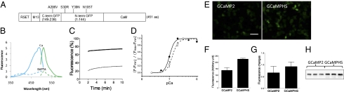
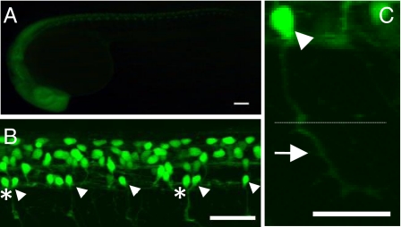
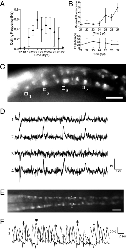
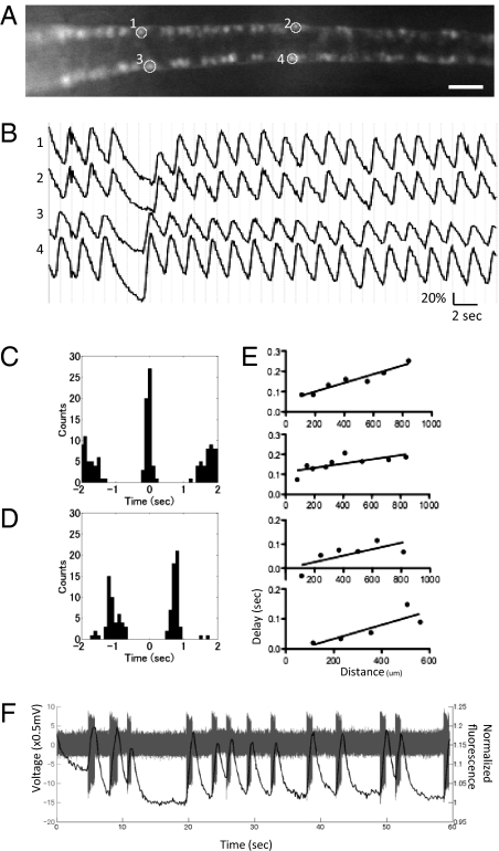
Animal behaviors are generated by well-coordinated activation of neural circuits. In zebrafish, embryos start to show spontaneous muscle contractions at 17 to 19 h postfertilization. To visualize how motor circuits in the spinal cord are activated during this behavior, we developed GCaMP-HS (GCaMP-hyper sensitive), an improved version of the genetically encoded calcium indicator GCaMP, and created transgenic zebrafish carrying the GCaMP-HS gene downstream of the Gal4-recognition sequence, UAS (upstream activation sequence). Then we performed a gene-trap screen and identified the SAIGFF213A transgenic fish that expressed Gal4FF, a modified version of Gal4, in a subset of spinal neurons including the caudal primary (CaP) motor neurons. We conducted calcium imaging using the SAIGFF213A; UAS:GCaMP-HS double transgenic embryos during the spontaneous contractions. We demonstrated periodic and synchronized activation of a set of ipsilateral motor neurons located on the right and left trunk in accordance with actual muscle movements. The synchronized activation of contralateral motor neurons occurred alternately with a regular interval. Furthermore, a detailed analysis revealed rostral-to-caudal propagation of activation of the ipsilateral motor neuron, which is similar to but much slower than the rostrocaudal delay observed during swimming in later stages. Our study thus demonstrated coordinated activities of the motor neurons during the first behavior in a vertebrate. We propose the GCaMP technology combined with the Gal4FF-UAS system is a powerful tool to study functional neural circuits in zebrafish.
Conflict of interest statement
The authors declare no conflict of interest.
Figures
Similar articles
-
Genetic dissection of neural circuits by Tol2 transposon-mediated Gal4 gene and enhancer trapping in zebrafish.Proc Natl Acad Sci U S A. 2008 Jan 29;105(4):1255-60. doi: 10.1073/pnas.0704963105. Epub 2008 Jan 17. Proc Natl Acad Sci U S A. 2008. PMID: 18202183 Free PMC article.
-
zTrap: zebrafish gene trap and enhancer trap database.BMC Dev Biol. 2010 Oct 18;10:105. doi: 10.1186/1471-213X-10-105. BMC Dev Biol. 2010. PMID: 20950494 Free PMC article.
-
A hybrid electrical/chemical circuit in the spinal cord generates a transient embryonic motor behavior.J Neurosci. 2014 Jul 16;34(29):9644-55. doi: 10.1523/JNEUROSCI.1225-14.2014. J Neurosci. 2014. PMID: 25031404 Free PMC article.
-
Gal4 Driver Transgenic Zebrafish: Powerful Tools to Study Developmental Biology, Organogenesis, and Neuroscience.Adv Genet. 2016;95:65-87. doi: 10.1016/bs.adgen.2016.04.002. Epub 2016 Jun 13. Adv Genet. 2016. PMID: 27503354 Review.
-
Imaging spinal neuron ensembles active during locomotion with genetically encoded calcium indicators.Ann N Y Acad Sci. 2013 Mar;1279:71-9. doi: 10.1111/nyas.12092. Ann N Y Acad Sci. 2013. PMID: 23531004 Free PMC article. Review.
Cited by
-
Biocompatibility of a genetically encoded calcium indicator in a transgenic mouse model.Nat Commun. 2012;3:1031. doi: 10.1038/ncomms2035. Nat Commun. 2012. PMID: 22929788
-
Generation of GCaMP6s-Expressing Zebrafish to Monitor Spatiotemporal Dynamics of Calcium Signaling Elicited by Heat Stress.Int J Mol Sci. 2021 May 24;22(11):5551. doi: 10.3390/ijms22115551. Int J Mol Sci. 2021. PMID: 34074030 Free PMC article.
-
Optical Recording of Action Potential Initiation and Propagation in Mouse Skeletal Muscle Fibers.Biophys J. 2018 Dec 4;115(11):2127-2140. doi: 10.1016/j.bpj.2018.10.026. Epub 2018 Nov 3. Biophys J. 2018. PMID: 30448039 Free PMC article.
-
A synaptic mechanism for network synchrony.Front Cell Neurosci. 2014 Sep 18;8:290. doi: 10.3389/fncel.2014.00290. eCollection 2014. Front Cell Neurosci. 2014. PMID: 25278839 Free PMC article. Review.
-
Convergence Circuit Mapping: Genetic Approaches From Structure to Function.Front Syst Neurosci. 2021 Jun 21;15:688673. doi: 10.3389/fnsys.2021.688673. eCollection 2021. Front Syst Neurosci. 2021. PMID: 34234652 Free PMC article. Review.
References
-
- Saint-Amant L, Drapeau P. Time course of the development of motor behaviors in the zebrafish embryo. J Neurobiol. 1998;37:622–632. - PubMed
-
- Saint-Amant L, Drapeau P. Synchronization of an embryonic network of identified spinal interneurons solely by electrical coupling. Neuron. 2001;31:1035–1046. - PubMed
-
- Fetcho JR, O'Malley DM. Visualization of active neural circuitry in the spinal cord of intact zebrafish. J Neurophysiol. 1995;73:399–406. - PubMed
-
- Smetters D, Majewska A, Yuste R. Detecting action potentials in neuronal populations with calcium imaging. Methods. 1999;18:215–221. - PubMed
Publication types
MeSH terms
Substances
LinkOut - more resources
Full Text Sources
Molecular Biology Databases
Research Materials
Miscellaneous
