. 2011 Mar 22;108(12):4892-6.
doi: 10.1073/pnas.1018202108. Epub 2011 Mar 7.

Most fertilizing mouse spermatozoa begin their acrosome reaction before contact with the zona pellucida during in vitro fertilization

Affiliations

Most fertilizing mouse spermatozoa begin their acrosome reaction before contact with the zona pellucida during in vitro fertilization

Mayuko Jin et al. Proc Natl Acad Sci U S A. .

Abstract

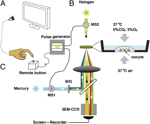
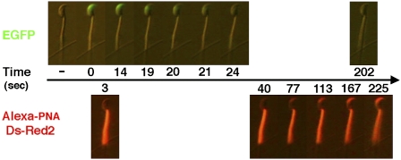
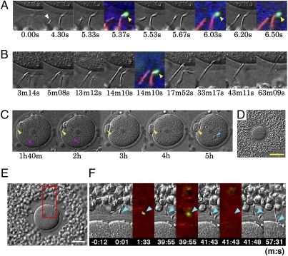
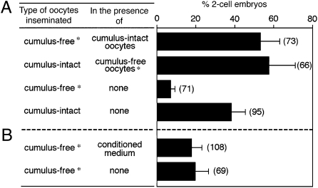
To fuse with oocytes, spermatozoa of eutherian mammals must pass through extracellular coats, the cumulus cell layer, and the zona pellucida (ZP). It is generally believed that the acrosome reaction (AR) of spermatozoa, essential for zona penetration and fusion with oocytes, is triggered by sperm contact with the zona pellucida. Therefore, in most previous studies of sperm-oocyte interactions in the mouse, the cumulus has been removed before insemination to facilitate the examination of sperm-zona interactions. We used transgenic mouse spermatozoa, which enabled us to detect the onset of the acrosome reaction using fluorescence microscopy. We found that the spermatozoa that began the acrosome reaction before reaching the zona were able to penetrate the zona and fused with the oocyte's plasma membrane. In fact, most fertilizing spermatozoa underwent the acrosome reaction before reaching the zona pellucida of cumulus-enclosed oocytes, at least under the experimental conditions we used. The incidence of in vitro fertilization of cumulus-free oocytes was increased by coincubating oocytes with cumulus cells, suggesting an important role for cumulus cells and their matrix in natural fertilization.


Conflict of interest statement

The authors declare no conflict of interest.

Figures

Fig. 1.
Fig. 1.
The video microscopic in vitro fertilization system consists of three components. (A) Oocyte-holding Petri dish (Materials and Methods). An oocyte surrounded by cumulus cell mass was slightly compressed under a 3 × 3 mm coverslip supported by four silicon grease spots. The dish was placed into an incubation chamber supplied with a mixed gas (5% CO2, 5% O2, and 90% N2). (B) Mercury–halogen illumination system: two sets of mechanical shutters (VS25; Uniblitz), one near the mercury lamp (MS1) and the other next to the halogen lamp (MS2), were controlled via a pulse generator. (C) Image-capturing system: a supersensitive video camera (NC-R550b; NEC) and a blue/green dual bandpass filter set enabled simultaneous visualization of EGFP and Ds-Red2 fluorescence in swimming spermatozoa. All spermatozoa entering the cumulus were viewed first at low magnification (10× objective lens). When a spermatozoon was seen approaching the oocyte, it was then focused under higher magnification (20× objective lens; Fig. S6). The spermatozoon under observation was exposed to blue/green light intermittently (125 ms) by pressing a remote shutter control button. After about 90 min of recording, all of the spermatozoa were individually analyzed for the status of their acrosomes. Motile (live) spermatozoa with EGFP fluorescence in their acrosomes were considered “acrosome intact” and those without EGFP fluorescence were considered “acrosome reacted.”
Fig. 2.
Fig. 2.
The progression of acrosomal exocytosis. Capacitated spermatozoa immobilized on a coverslip were incubated with BSA–HTF containing Alexa-594 PNA and thereafter observed under an epifluorescence microscope. Representative time-lapse photographs show a spermatozoon undergoing the acrosomal exocytosis induced by 10 μM ionomycin. Acrosomal EGFP disappearance took place within about 10 s followed by acrosomal staining with PNA lectin.
Fig. 3.
Fig. 3.
Viewing a fertilizing spermatozoon. (A–D) Photographs showing sperm penetration into the ZP and the cytoplasm of a single oocyte. Whether it was the actual fertilizing spermatozoon was determined retrospectively after video recording. Times are shown in seconds (s), minutes (m), and hours (h) after the fertilizing spermatozoon entered the field of view (0 s). (A) Successive stages of sperm attachment to the ZP. The fertilizing spermatozoon (arrowhead at 4.30 s) had no EGPF fluorescence (i.e., was acrosome reacted) before reaching the ZP surface at 6.20 s. (B) The head of this fertilizing spermatozoon passed through the ZP and reached the egg surface at 13 m 12 s. Note that an acrosome-intact spermatozoon (arrowhead at 14 m 10 s) with EGFP fluorescence remained on the ZP surface. (C) The oocyte under low magnification, showing formation of a second polar body (yellow arrowheads), sperm tail (red arrowheads), and sperm pronucleus (blue arrowhead). (D) The cumulus-enclosed oocyte before insemination under low magnification. (Scale bar, 100 μm.) (E and F) Fertilization by a spermatozoon that had undergone the AR after binding to the ZP. (E) A low-magnification view of a cumulus-enclosed oocyte used in this experiment. (Scale bar, 50 μm.) (F) The time of initial sperm binding to the ZP was set at 0. This spermatozoon remained on the ZP surface with an intact acrosome until 39 m 55 s and thereafter lost acrosomal EGFP by the time of the next fluorescence exposure at 41 m 43 s. After the AR, this spermatozoon initiated ZP penetration and fused with the plasma membrane of the oocyte at about 57 m 31 s. Arrowheads point to the sperm head.
Fig. 4.
Fig. 4.
The presence of an intact cumulus layer on oocytes increases the fertilizing ability of capacitated spermatozoa. Asterisks denote cumulus-free oocytes from transgenic females whose oocytes express red fluorescence. (A and B) Approximately 10–20 cumulus-free oocytes incubated with or without cumulus-enclosed oocytes (A) or with cumulus–oocyte complex conditioned medium (B) were inseminated at a final concentration of 2 × 10⁴ sperm/mL. The numbers of two-cell embryos per total oocytes examined was scored after 24 h. Shown are the percentages obtained from seven independent experiments. The total number of oocytes scored is indicated in parentheses.

Comment in

References

    1. Yanagimachi R. Mammalian fertilization. In: Knobil E, Neil J, editors. The Physiology of Reproduction. New York: Raven; 1994. pp. 189–317.
    1. Liu C, et al. Targeted disruption of the mZP3 gene results in production of eggs lacking a zona pellucida and infertility in female mice. Proc Natl Acad Sci USA. 1996;93:5431–5436. - PMC - PubMed
    1. Rankin T, et al. Mice homozygous for an insertional mutation in the Zp3 gene lack a zona pellucida and are infertile. Development. 1996;122:2903–2910. - PubMed
    1. Hoodbhoy T, Dean J. Insights into the molecular basis of sperm-egg recognition in mammals. Reproduction. 2004;127:417–422. - PubMed
    1. Gahlay G, Gauthier L, Baibakov B, Epifano O, Dean J. Gamete recognition in mice depends on the cleavage status of an egg's zona pellucida protein. Science. 2010;329:216–219. - PMC - PubMed

Publication types