. 2011 Mar 22;108(12):4840-5.
doi: 10.1073/pnas.1101734108. Epub 2011 Mar 7.

Mutated beta-catenin evades a microRNA-dependent regulatory loop

Affiliations

Mutated beta-catenin evades a microRNA-dependent regulatory loop

Angelo Veronese et al. Proc Natl Acad Sci U S A. .

Abstract

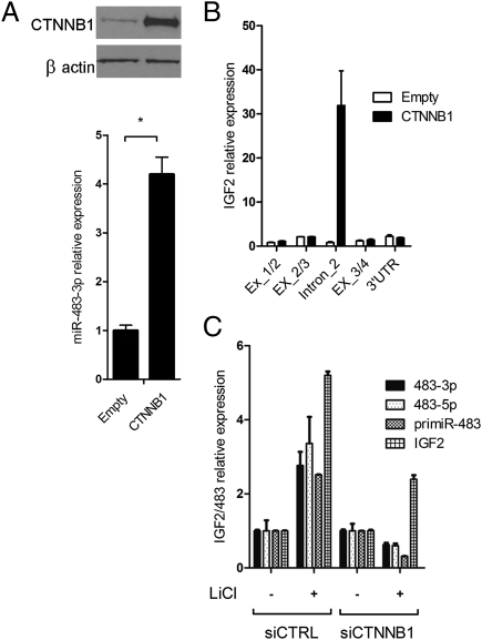
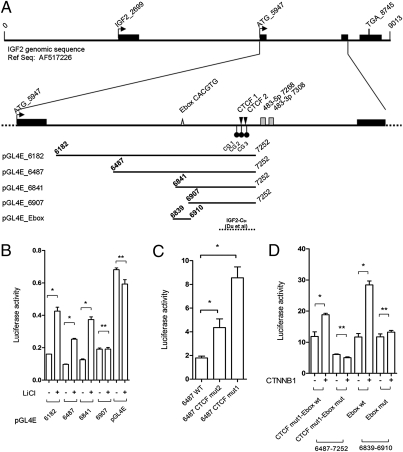
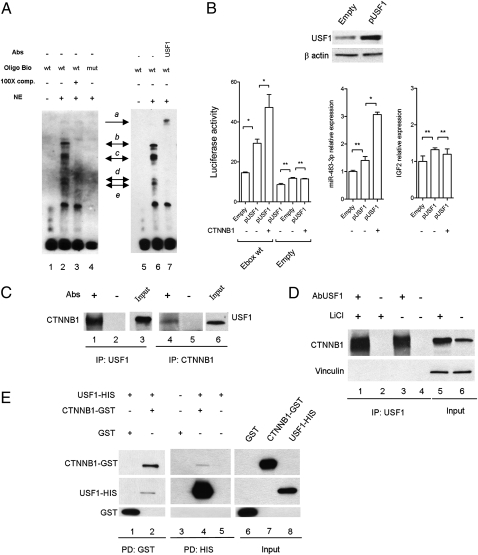
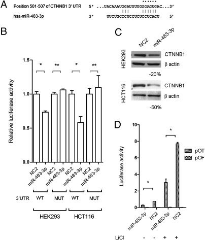
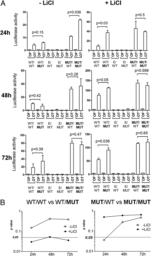
hsa-mir-483 is located within intron 2 of the IGF2 gene. We have previously shown oncogenic features of miR-483-3p through cooperation with IGF2 or by independently targeting the proapoptotic gene BBC3/PUMA. Here we demonstrate that expression of miR-483 can be induced independently of IGF2 by the oncoprotein β-catenin through an interaction with the basic helix-loop-helix protein upstream stimulatory transcription factor 1. We also show that β-catenin itself is a target of miR-483-3p, triggering a negative regulatory loop that becomes ineffective in cells harboring an activating mutation of β-catenin. These results provide insights into the complex regulation of the IGF2/miR-483 locus, revealing players in the β-catenin pathway.


Conflict of interest statement

The authors declare no conflict of interest.

Figures

Fig. 1.
Fig. 1.
Induction of miR-483-3p expression by β-catenin. (A) miR-483-3p was induced by enforced expression of wild-type CTNNB1. Expression value was related to miRNA expression on empty vector transfected cells (2−ΔΔCt). The expression of CTNNB1 was assessed by Western blot (Upper). *P < 0.02. (B) Expression analyses across the IGF2 locus modulated by the enforced expression of β-catenin. Three PCR products were designed to amplify IGF2 exon junctions (Ex_1/2, Ex_2/3, and Ex_3/4) and one its 3′UTR. One set of primers was located within intron 2 and was used to assess the expression of miR-483 precursor (intron_2). qRT-PCR was carried out with SYBR Green technologies. RNA was previously treated with DNase to avoid genomic contamination. Black bars indicate the expression detected in cells transfected with the CTNNB1-expressing vector; white bars indicate the expression detected in cells transfected with the empty vector. (C) Expression analysis of miR-483-3p, miR-483-5p, pri-miR-483, and IGF2 genes assayed by qRT-PCR using TaqMan probes after LiCl treatment (20 mM for 24 h) with and without siRNA for CTNNB1 gene. IGF2 expression was still induced, although more weakly than the control. This IGF2 induction by LiCl has not been previously described, and is not necessarily due to β-catenin because of the large number of pathways affected by GSK3B inhibition.
Fig. 2.
Fig. 2.
Analysis of the miR-483 minimal promoter region responsive to LiCl/CTNNB1 stimuli. (A) Genomic structure of the IGF2/483 locus from the reference AF517226 genomic sequence. Exons (black bars), start (ATG_5947) and stop codons (TGA_8745), E-box elements (gray triangle), the two predicted CTCF binding sites (black triangle), three CpG dinucleotides around the CTCF binding sites (black circles), and miR-483-3p and miR-483-5p (gray boxes) are shown. The five genomic fragments cloned upstream of the luciferase reporter gene in pGL4E for the analysis of the promoter are indicated at the bottom of the panel. The genomic region with insulator activity studied by Du et al. (22) is indicated (broken line). (B) Luciferase activity of four genomic fragments cloned in pGL4E with and without LiCl treatment; the pGL4E empty vector was used as control. (C) Luciferase activity of the wild-type pGL4E-6487 (WT) and the mutated pGL4E-6487 in the predicted CTCF binding sites (CTCF mut1 and CTCF mut2). (D) Analysis of the wild-type E-box element (Ebox wt) and mutant (Ebox mut) by luciferase assay of the pGL4E-6487 CTCF mut1 and pGL4E_Ebox. Firefly luciferase activity was normalized on Renilla luciferase activity of the cotransfected pGL4R vector. *P < 0.02, **P > 0.02.
Fig. 3.
Fig. 3.
USF1 serves as a mediator between β-catenin and the miR-483 locus. (A) EMSA of nuclear extract (NE) from HEK293 cells using the miR-483 E-box probe (lanes 1–3 and 5–7) or the mutant form (lane 4). The specific complexes are indicated by black arrows (be). Lane 7 shows the supershift generated by USF1 antibody (complex a). (B) Luciferase assay of wild-type pGL4E_E box after enforced expression of USF1 and CTNNB1 using the pGL4E empty vector as control (Left). The center and right panels show the expression of miR-483-3p and IGF2 gene, respectively, after enforced expression of USF1 and CTNNB1. The miR-483-3p and IGF2 expression values of the empty vector were the controls. The exogenous expression of USF1 was assessed by Western blot (Upper). *P < 0.02, **P > 0.02. (C) Nuclear extract from HEK293 cells were immunoprecipitated with either USF1 (lanes 1–3) or CTNNB1 antibodies (lanes 4–6). After being washed, samples were run on an SDS/PAGE gel and transferred to nitrocellulose. The blots were probed with USF1 and CTNNB1 antibodies. (D) Nuclear extract from HEK293 cells with (lanes 1 and 2) and without (lanes 3 and 4) treatment with LiCl (20 mM for 24 h) was immunoprecipitated with USF1 antibody and the blot was probed for CTNNB1 protein. The input shows an incremented quantity of CTNNB1 after LiCl treatment (lanes 5 and 6). Vinculin protein expression was used as loading protein control. (E) GST-CTNNB1 and HIS-USF1 fusion proteins were subjected to GST (lanes 1 and 2) and HIS (lanes 3–5) pull-down analysis. Binding reaction products were washed, and proteins were separated by SDS/PAGE. The membrane was probed with anti-CTNNB1, anti-USF1, and GST antibodies.
Fig. 4.
Fig. 4.
β-Catenin is a target of miR-483-3p. (A) Putative binding site of miR-483-3p in CTNNB1 3′UTRs (TargetScan database). Asterisks indicate nucleotides substituted in 3′UTR miR-483-3p predicted target site to perform luciferase assay. (B) CTNNB1 3′UTRs regulate luciferase activity dependent on miR-483-3p in HEK293 and HCT116 cell lines. MUT, mutant; WT, wild type. (C) Western blot analysis of CTNNB1 after miR-483-3p transfection in HEK293 and HCT116 cell lines. Cells were collected 48 h after miRNA transfection. (D) Luciferase activity of the reporter vectors pOT and pOF in HCT116 cells cotransfected with miR-483-3p and scramble oligo (NC2) with and without LiCl treatment. *P < 0.02, **P > 0.02.
Fig. 5.
Fig. 5.
Mutated form of β-catenin evades the miR-483-3p regulatory loop. (A) Luciferase activity during the time (24, 48, and 72 h) of the reporter vectors pOT and pOF in HEK293 cells cotransfected with the four different CTNNB1 expression vectors (mutational status of the CDS and 3′UTR miR-483-3p target site of CTNNB1: CDS/3′UTR) with (Right) and without LiCl treatment (Left). As experiment controls, wild-type CTNNB1 3′UTR and mutated mir-483-3p target were cloned in the expression vector pIRESNeo2. E/MUT, empty/MUT; E/WT, empty/WT. (B) P values are indicated and graphically represented.

References

    1. Perez-Moreno M, Jamora C, Fuchs E. Sticky business: Orchestrating cellular signals at adherens junctions. Cell. 2003;112:535–548. - PubMed
    1. Clevers H. Wnt/β-catenin signaling in development and disease. Cell. 2006;127:469–480. - PubMed
    1. Wang SS, et al. Alterations of the PPP2R1B gene in human lung and colon cancer. Science. 1998;282:284–287. - PubMed
    1. Satoh S, et al. AXIN1 mutations in hepatocellular carcinomas, and growth suppression in cancer cells by virus-mediated transfer of AXIN1. Nat Genet. 2000;24:245–250. - PubMed
    1. Major MB, et al. Wilms tumor suppressor WTX negatively regulates WNT/β-catenin signaling. Science. 2007;316:1043–1046. - PubMed

Publication types

MeSH terms

LinkOut - more resources