. 2011 Mar 22;108(12):4962-7.
doi: 10.1073/pnas.1016005108. Epub 2011 Mar 7.

Inflammation-dependent secretion and splicing of IL-32{gamma} in rheumatoid arthritis

Affiliations

Inflammation-dependent secretion and splicing of IL-32{gamma} in rheumatoid arthritis

Bas Heinhuis et al. Proc Natl Acad Sci U S A. .

Abstract

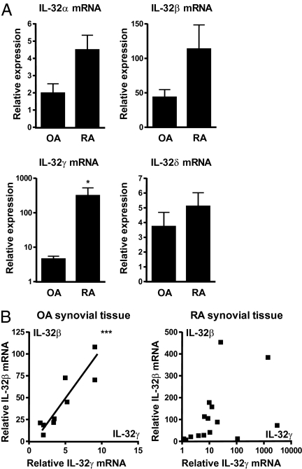
Different splice variants of the proinflammatory cytokine IL-32 are found in various tissues; their putative differences in biological function remain unknown. In the present study, we report that IL-32γ is the most active isoform of the cytokine. Splicing to one less active IL-32β appears to be a salvage mechanism to reduce inflammation. Adenoviral overexpression of IL-32γ (AdIL-32γ) resulted in exclusion of the IL-32γ-specific exon in vitro as well as in vivo, primarily leading to expression of IL-32β mRNA and protein. Splicing of the IL-32γ-specific exon was prevented by single-nucleotide mutation, which blocked recognition of the splice site by the spliceosome. Overexpression of splice-resistant IL-32γ in THP1 cells or rheumatoid arthritis (RA) synovial fibroblasts resulted in a greater induction of proinflammatory cytokines such as IL-1β, compared with IL-32β. Intraarticular introduction of IL-32γ in mice resulted in joint inflammation and induction of several mediators associated with joint destruction. In RA synovial fibroblasts, overexpression of primarily IL-32β showed minimal secretion and reduced cytokine production. In contrast, overexpression of splice-resistant IL-32γ in RA synovial fibroblasts exhibited marked secretion of IL-32γ. In RA, we observed increased IL-32γ expression compared with osteoarthritis synovial tissue. Furthermore, expression of TNFα and IL-6 correlated significantly with IL-32γ expression in RA, whereas this was not observed for IL-32β. These data reveal that naturally occurring IL-32γ can be spliced into IL-32β, which is a less potent proinflammatory mediator. Splicing of IL-32γ into IL-32β is a safety switch in controlling the effects of IL-32γ and thereby reduces chronic inflammation.


Conflict of interest statement

The authors declare no conflict of interest.

Figures

Fig. 1.
Fig. 1.
IL-32α, IL-32β, IL-32γ, and IL-32δ expression and correlation in OA and RA synovial biopsies. RNA from synovial biopsies was isolated and used for determining IL-32 isoform expression. (A) IL-32γ mRNA expression was significantly enhanced in RA versus OA synovial biopsies (mean ± SEM; OA, n = 9; RA, n = 15; Mann–Whitney U test; *P < 0.05). (B) Correlation between IL-32γ and IL-32β isoforms in OA and RA synovial tissue (OA, n = 9, Pearson correlation test, Pearson r = 0.9023, P = 0.0009, ***P < 0.001; RA, n = 15, Pearson correlation test, nonsignificant).
Fig. 2.
Fig. 2.
Correlation between IL-32γ and TNFα or IL-6 in RA synovial tissue. (A) Enhanced TNFα, IL-1β, IL-6, and CXCL8 expression in RA synovial tissue (mean ± SEM; OA, n = 9; RA, n = 14; Mann-Whitney U test). (B) TNFα and IL-6 both correlated with IL-32γ expression in RA synovial tissue (RA, n = 14, Pearson correlation test, IL-6/IL-32γ, Pearson r = 0.9638, P < 0.0001; TNFα/IL-32γ, Pearson r = 0.9598, P < 0.0001, ***P < 0.001).
Fig. 3.
Fig. 3.
Splicing of IL-32γ results in expression of IL-32β, which can be prevented by single-nucleotide mutation. (A) Adenoviral overexpression of IL-32γ resulted in IL-32β and IL-32γ mRNA expression and primarily IL-32β protein production, as shown in HeLa cells. (B) Theoretical model. G-to-A mutation of the B2 donor splice site prevents IL-32γ-into-IL-32β splicing. (C) Overexpression of IL-32γM results in IL-32γ mRNA expression and protein production in HeLa cells. Furthermore, in THP1 cells, AdControl shows no IL-32 protein production, whereas AdIL-32γ–exposed cells show primarily IL-32β and some IL-32γ production. Moreover, AdIL-32γM-transduced cells show primarily IL-32γ and some IL-32β production. In addition, we quantified IL-32β or IL-32γ production and corrected for actin production in AdIL-32γ– or AdIL-32γM–transduced cells, respectively. (D) THP1 cells transduced with AdControl, AdIL-32γ, or AdIL-32γM followed by medium or IL-1β stimulation. AdIL-32γM-transduced THP1 cells showed enhanced secretion of IL-6 and CXCL8 protein compared with AdIL-32γ– or AdControl-exposed cells. Furthermore, AdIL-32γM-exposed cells showed enhanced IL-1β production compared with AdControl-transduced cells. Additionally, AdIL-32γ showed only enhanced CXCL8 production compared with AdControl-transduced cells (mean ± SEM, n = 4, Bonferroni's multiple-comparison test, *P < 0.05, ***P < 0.001).
Fig. 4.
Fig. 4.
Aggravated arthritis in AdIL-32γM-exposed mice. (A) Splicing of IL-32γ into IL-32β in 3T3 cells and mice, which is not observed in AdIL-32γM-exposed mice. Enhanced macroscopic knee joint scores (mean ± SEM, n = 12, Mann–Whitney U test, ***P < 0.001) and more infiltrating cells in mice knee joints (mean ± SEM, n = 6, Mann–Whitney U test, **P < 0.01), which is shown by H&E staining. (B) Increased expression of TNFα, IL-1β, IL-6, and IL-32γ (1 × 5 pooled synovial biopsies), whereas IL-32all expression was comparable between AdIL-32γ and AdIL-32γM. (C) Enhanced iNOS, MMP3, and MMP13 expression in patellar cartilage derived from AdIL-32γM-exposed mice (1 × 5 pooled patellar cartilage samples).
Fig. 5.
Fig. 5.
Secretion of IL-32 can be enhanced by IL-1β or TNFα stimulation, whereas splice-resistant IL-32γ is directly secreted in RA synovial fibroblasts. (A) AdControl-transduced RA FLS showed low IL-32 concentrations both intra- and extracellularly. Secretion of IL-32 was significantly induced in AdIL-32γ–transduced RA FLS after IL-1β or TNFα stimulation, whereas medium control showed minimal secretion of IL-32 (mean ± SEM, n = 5, Mann–Whitney U test, *P < 0.05, **P < 0.01). Moreover, AdIL-32γM-transduced RA FLS showed impressive secretion of IL-32γ compared with AdIL-32γ, whereas IL-1β or TNFα stimulation showed some increase (mean ± SEM, n = 5, Mann–Whitney U test, *P < 0.05, **P < 0.01). (B) In addition, cell death was investigated by determining LDH release, which was not different between the groups (mean percentage LDH release ± SEM, n = 6, Dunnett's multiple-comparison test). (C) Splice-resistant IL-32γ was more potent in inducing IL-6 and CXCL8 compared with spliced IL-32γ or AdControl after IL-1β stimulation (fold increase protein level ± SEM, n = 5, Bonferroni's multiple-comparison test, *P < 0.05, **P < 0.01).

References

    1. Goda C, et al. Involvement of IL-32 in activation-induced cell death in T cells. Int Immunol. 2006;18:233–240. - PubMed
    1. Kim SH, Han SY, Azam T, Yoon DY, Dinarello CA. Interleukin-32: A cytokine and inducer of TNFα. Immunity. 2005;22:131–142. - PubMed
    1. Dahl CA, Schall RP, He HL, Cairns JS. Identification of a novel gene expressed in activated natural killer cells and T cells. J Immunol. 1992;148:597–603. - PubMed
    1. Mun SH, et al. Tumor necrosis factor α-induced interleukin-32 is positively regulated via the Syk/protein kinase Cδ/JNK pathway in rheumatoid synovial fibroblasts. Arthritis Rheum. 2009;60:678–685. - PubMed
    1. Shioya M, et al. Epithelial overexpression of interleukin-32α in inflammatory bowel disease. Clin Exp Immunol. 2007;149:480–486. - PMC - PubMed

Publication types

MeSH terms