. 2011 Apr;21(4):535-44.
doi: 10.1101/gr.114579.110. Epub 2011 Mar 7.

Recurrence, submicroscopic complexity, and potential clinical relevance of copy gains detected by array CGH that are shown to be unbalanced insertions by FISH

Affiliations

Recurrence, submicroscopic complexity, and potential clinical relevance of copy gains detected by array CGH that are shown to be unbalanced insertions by FISH

Nicholas J Neill et al. Genome Res. 2011 Apr.

Abstract

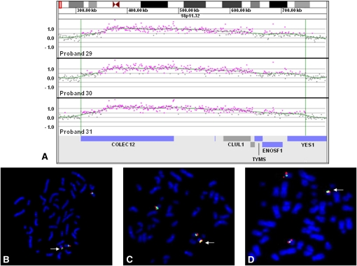
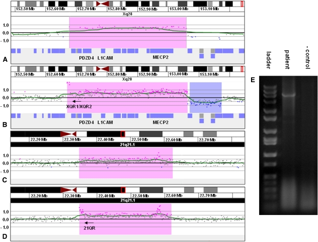
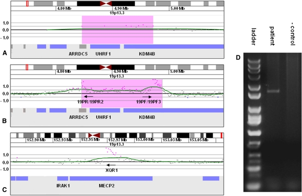
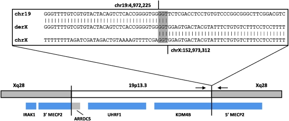
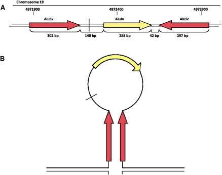
Insertions occur when a segment of one chromosome is translocated and inserted into a new region of the same chromosome or a non-homologous chromosome. We report 71 cases with unbalanced insertions identified using array CGH and FISH in 4909 cases referred to our laboratory for array CGH and found to have copy-number abnormalities. Although the majority of insertions were non-recurrent, several recurrent unbalanced insertions were detected, including three der(Y)ins(Y;18)(q?11.2;p11.32p11.32)pat inherited from parents carrying an unbalanced insertion. The clinical significance of these recurrent rearrangements is unclear, although the small size, limited gene content, and inheritance pattern of each suggests that the phenotypic consequences may be benign. Cryptic, submicroscopic duplications were observed at or near the insertion sites in two patients, further confounding the clinical interpretation of these insertions. Using FISH, linear amplification, and array CGH, we identified a 126-kb duplicated region from 19p13.3 inserted into MECP2 at Xq28 in a patient with symptoms of Rett syndrome. Our results demonstrate that although the interpretation of most non-recurrent insertions is unclear without high-resolution insertion site characterization, the potential for an otherwise benign duplication to result in a clinically relevant outcome through the disruption of a gene necessitates the use of FISH to determine whether copy-number gains detected by array CGH represent tandem duplications or unbalanced insertions. Further follow-up testing using techniques such as linear amplification or sequencing should be used to determine gene involvement at the insertion site after FISH has identified the presence of an insertion.


Figures

Figure 1.
Figure 1.
Characterization of recurrent der(Y)ins(Y;18)(?p11.2;p11.32p11.32) by oligonucleotide array CGH and FISH. (A) Oligonucleotide microarray results showing identical single-copy gains of 340 probes from 18p11.32, ∼437 kb in size (chr18:309,785–747,102 based on UCSC 2006 hg 18 assembly), in three probands. Probes are ordered on the x-axis according to physical mapping positions, with the most distal 18p11.32 probes to the left and the most proximal 18p11.32 probes to the right. Values along the y-axis represent log2 ratios of patient:control signal intensities. Genes in the interval are shown as blue and gray bars below. (B) Metaphase FISH results showing insertion of RP11-720L2 (red) from the duplicated region on chromosome 18 in proband 29 into the pericentromeric region of the Y chromosome (arrow). (Green) A centromere probe for chromosome 18 and a Yp11.31 probe specific to SRY (RP11-400O10). (C) Metaphase FISH results showing insertion of RP11-133D9 (red) from the duplicated region on chromosome 18 into the pericentromeric region of the Y chromosome in proband 30 (arrow). (Green) Centromere probes for chromosome 18 and the Y chromosome. (D) Metaphase FISH results showing the insertion of RP11-720L2 (red) into the pericentromeric region of the Y chromosome in proband 31 (arrow). (Green) Centromere probes for chromosome 18 and the Y chromosome.
Figure 2.
Figure 2.
Characterization of der(21)ins(21;X)(q21.1;q28q28)dup(21)(q21.1q21.1) by oligonucleotide array CGH, linear amplification, and PCR. (A) 135k feature oligonucleotide microarray results showing a single-copy gain of 48 probes, ∼382 kb in size (chrX:152,676,843–153,058,941 based on the UCSC 2006 hg18 assembly), from Xq28 in proband 2. Probes are ordered on the x-axis with the most proximal Xq28 probes to the left and the most distal Xq28 probes to the right. Values along the y-axis represent log2 ratios of patient:control signal intensities. (B) 2.1M feature oligonucleotide microarray results showing the same duplication as in A after linear amplification with primers XQR1 and XQR2. Successful amplification is evidenced by the elevated log ratios of probes in the proximal portion of the duplicated region. (C) 2.1M feature oligonucleotide microarray results showing a single-copy gain of 210 probes, ∼272 kb in size (chr21:22,347,877–22,623,043 based on the UCSC 2006 hg18 assembly), from 21q21.1 in proband 2. (D) 2.1 M feature oligonucleotide microarray results showing the same duplication as in C after linear amplification with the primers from B. The elevated log ratios in the proximal portion of the duplicated segment are indicative of the insertion of Xq22.2 sequence into 21q21.1, which allowed for continuous amplification across the breakpoint. The cluster of elevated probes that can be seen in the distal portion of the duplicated segment was also present in the unamplified sample and probably represents a CNV or artifact. (E) Gel electrophoresis of PCR amplicon produced with primers XQR1 and 21QR confirming the insertion site detected by linear amplification.
Figure 3.
Figure 3.
Characterization of der(X)ins(X;19)(q28;p13.3p13.3) by oligonucleotide array CGH, linear amplification, and PCR. (A) 135k-feature oligonucleotide microarray results showing a single-copy gain of 9 probes, ∼126 kb in size (chr19:4,845,920–4,971,768 based on the UCSC 2006 hg 18 assembly), from 19p13.3 in proband 6. Probes are ordered along the x-axis with the most distal 19p13.3 probes to the left and the most proximal 19p13.3 probes to the right. Values along the y-axis represent log2 ratios of patient:control signal intensities. (B) 2.1M feature oligonucleotide microarray analysis showing the same duplication as in A after linear amplification with primers 19PF, 19PF3, 19PR, and 19PR2. Successful amplification is evidenced by the elevated log ratios of probes at the proximal and distal edges of the duplicated region. (C) 2.1M feature oligonucleotide microarray analysis showing linear amplification from B extending across the insertion junction into intron 2 of MECP2. (D) Gel electrophoresis of PCR amplicon produced with primers 19PF and XQR1 confirming the insertion site detected by linear amplification.
Figure 4.
Figure 4.
Diagram representing the insertion site in the der(X) resulting from insertion of chromosome 19 material into Xq28 in proband 6. (Gray shaded area within the inset box) The 3-nucleotide span of microhomology shared at the distal insertion breakpoint. (Small black arrows) The positions of primers used to generate the PCR fragment and obtain the junction sequence. Genes are displayed as blue and gray bars below, and show the disruption of MECP2 as well as the two possible fusion gene products that could result from the insertion.
Figure 5.
Figure 5.
(A) Diagram outlining the repetitive sequences around the proximal breakpoint of the copy-number gain on chromosome 19. (Vertical black line) The position of the insertion breakpoint. Members of AluS families (red boxes) and AluJ families (yellow boxes) of repetitive elements. (B) Diagram of potential stem–loop structure mediated by inverted AluS repeats present at the breakpoint.

References

    1. Abuelo DN, Barsel-Bowers G, Richardson A 1988. Insertional translocations: Report of two new families and review of the literature. Am J Med Genet 31: 319–329 - PubMed
    1. Ballif BC, Wakui K, Gajecka M, Shaffer LG 2004. Translocation breakpoint mapping and sequence analysis in three monosomy 1p36 subjects with der(1)t(1;1)(p36;q44) suggest mechanisms for telomere capture in stabilizing de novo terminal rearrangements. Hum Genet 114: 198–206 - PubMed
    1. Ballif BC, Theisen A, Coppinger J, Gowans GC, Hersh JH, Madan-Khetarpal S, Schmidt KR, Tervo R, Escobar LF, Friedrich CA, et al. 2008a. Expanding the clinical phenotype of the 3q29 microdeletion syndrome and characterization of the reciprocal microduplication. Mol Cytogenet 1: 8 doi: 10.1186/1755-8166-1-8 - PMC - PubMed
    1. Ballif BC, Theisen A, McDonald-McGinn DM, Zackai EH, Hersh JH, Bejjani BA, Shaffer LG 2008b. Identification of a previously unrecognized microdeletion syndrome of 16q11.2q12.2. Clin Genet 74: 469–475 - PubMed
    1. Baptista J, Prigmore E, Gribble SM, Jacobs PA, Carter NP, Crolla JA 2005. Molecular cytogenetic analyses of breakpoints in apparently balanced reciprocal translocations carried by phenotypically normal individuals. Eur J Hum Genet 13: 1205–1212 - PubMed