Review
. 2011 Mar;8(3):141-50.
doi: 10.1038/nrgastro.2011.2.

Cystic precursors to invasive pancreatic cancer

Affiliations
Review

Cystic precursors to invasive pancreatic cancer

Hanno Matthaei et al. Nat Rev Gastroenterol Hepatol. 2011 Mar.

Abstract

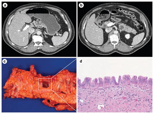
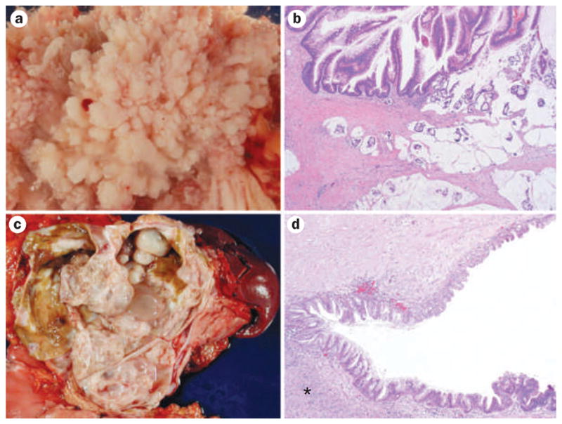
Improvements in the sensitivity and quality of cross-sectional imaging have led to increasing numbers of patients being diagnosed with cystic lesions of the pancreas. In parallel, clinical, radiological, pathological and molecular studies have improved the systems for classifying these cysts. Patients with asymptomatic serous cystic neoplasms can be managed conservatively with regular monitoring; however, the clinical management of patients with intraductal papillary mucinous neoplasms and mucinous cystic neoplasms is far more challenging, as it is difficult to determine whether these lesions will progress to malignancy. Fortunately, prospective studies have helped to establish that proposed clinical and radiological criteria (the Sendai guidelines) can be used to guide the care of patients with cystic lesions of the pancreas. Despite this progress in imaging and clinical guidelines, sensitive and specific tests have not yet been developed that can reliably predict the histology and biological properties of a cystic lesion. Such biomarkers are urgently needed, as noninvasive precursors of pancreatic cancer are curable, while the vast majority of invasive pancreatic adenocarcinomas are not.


Conflict of interest statement

Competing interests

The authors, the journal Chief Editor N. Wood and the CME questions author C. P. Vega declare no competing interests.

Figures

Figure 1
Figure 1
Multifocal intraductal papillary mucinous neoplasms (IPMNs) within a pancreas. CT imaging before surgery reveals multicystic changes involving the a | body of the pancreas and b | tail of the pancreas. c | After distal pancreatectomy six independent branch duct IPMNs can grossly be identified. d | Histologically the lesions show low to intermediate grade dysplasia and a characteristic gastric foveolar epithelial subtype (hematoxylin and eosin staining, 100× original magnification).
Figure 2
Figure 2
Cystic precursor lesions of pancreatic cancer. a | An extensive intraductal papillary growth of a large intraductal papillary mucinous neoplasm (IPMN) (image taken with specimen immersed in water). b | Histology displays finger-like papillae and high-grade dysplasia (upper part of image) with an invasive colloid adenocarinoma (lower part of image); these lesions are often located in the main pancreatic duct and show characteristic intestinal differentiation (hematoxylin and eosin staining, 20× original magnification). c | A large multicystic mucinous cystic neoplasm (MCN) in the pancreatic tail. d | A diagnostic sign for MCN ovarian-like stroma (indicated with an asterisk) underlies the neoplastic epithelium. The latter often shows abrupt transition in the degree of dysplasia; note the flat epithelium with mild cytoarchitectural atypia at the upper part of the image versus the more dysplastic and papillary growth pattern in the lower part of the image (hematoxylin and eosin staining, 40× original magnification).

References

    1. Brugge WR, et al. Diagnosis of pancreatic cystic neoplasms: a report of the cooperative pancreatic cyst study. Gastroenterology. 2004;126:1330–1336. - PubMed
    1. Fernández-del Castillo C, et al. Incidental pancreatic cysts: clinicopathologic characteristics and comparison with symptomatic patients. Arch Surg. 2003;138:427–434. - PMC - PubMed
    1. Spinelli KS, et al. Cystic pancreatic neoplasms: observe or operate. Ann Surg. 2004;239:651–659. - PMC - PubMed
    1. Winter JM, et al. Periampullary and pancreatic incidentaloma: a single institution’s experience with an increasingly common diagnosis. Ann Surg. 2006;243:673–683. - PMC - PubMed
    1. Yachida S, et al. Distant metastasis occurs late during the genetic evolution of pancreatic cancer. Nature. 2010;467:1114–1117. - PMC - PubMed

Publication types

MeSH terms