. 2011 May;91(5):665-74.
doi: 10.1038/labinvest.2011.7. Epub 2011 Mar 7.

Arterial pathology in canine mucopolysaccharidosis-I and response to therapy

Affiliations

Arterial pathology in canine mucopolysaccharidosis-I and response to therapy

Jeremiah A Lyons et al. Lab Invest. 2011 May.

Abstract

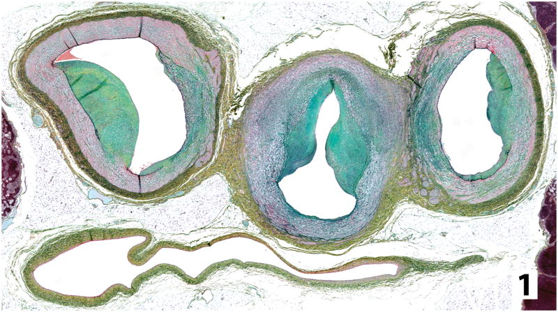
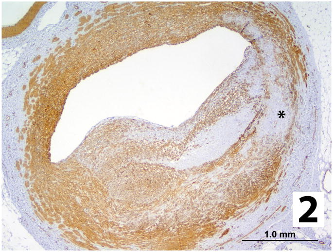
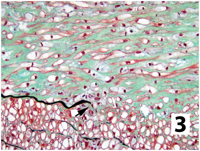
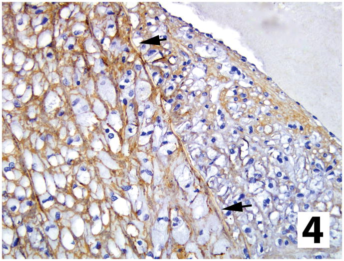
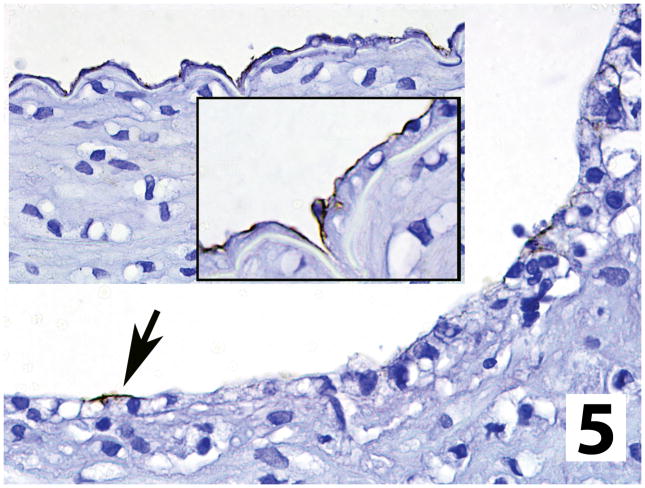
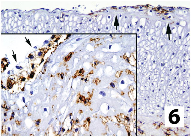
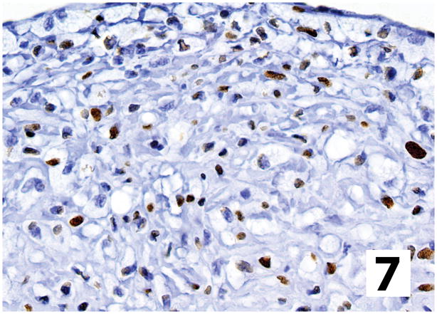
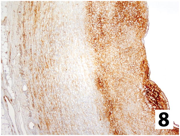
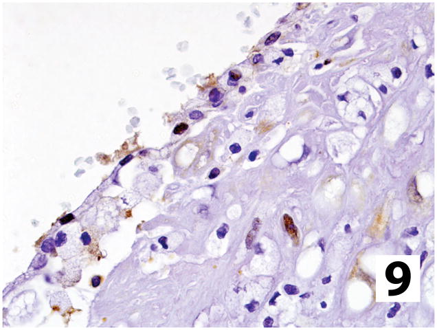
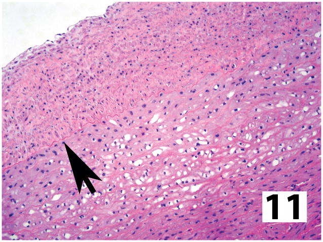
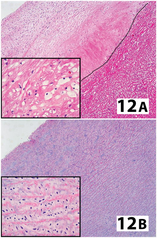
Mucopolysaccharidosis-I (MPS-I) is an inherited deficiency of α-L-iduronidase (IdU) that causes lysosomal accumulation of glycosaminoglycans (GAG) in a variety of parenchymal cell types and connective tissues. The fundamental link between genetic mutation and tissue GAG accumulation is clear, but relatively little attention has been given to the morphology or pathogenesis of associated lesions, particularly those affecting the vascular system. The terminal parietal branches of the abdominal aorta were examined from a colony of dogs homozygous (MPS-I affected) or heterozygous (unaffected carrier) for an IdU mutation that eliminated all enzyme activity, and in affected animals treated with human recombinant IdU. High-resolution computed tomography showed that vascular wall thickenings occurred in affected animals near branch points, and associated with low endothelial shear stress. Histologically these asymmetric 'plaques' entailed extensive intimal thickening with disruption of the internal elastic lamina, occluding more than 50% of the vascular lumen in some cases. Immunohistochemistry was used to show that areas of sclerosis contained foamy (GAG laden) macrophages, fibroblasts and smooth muscle cells, with loss of overlying endothelial basement membrane and claudin-5 expression. Lesions contained scattered cells expressing nuclear factor-κβ (p65), increased fibronectin and transforming growth factor β-1 signaling (with nuclear Smad3 accumulation) in comparison to unaffected vessels. Intimal lesion development and morphology was improved by intravenous recombinant enzyme treatment, particularly with immune tolerance to this exogenous protein. The progressive sclerotic vasculopathy of MPS-I shares some morphological and molecular similarities to atherosclerosis, including formation in areas of low shear stress near branch points, and can be reduced or inhibited by intravenous administration of recombinant IdU.


Conflict of interest statement

Conflict of interest: none declared.

Figures

Figure 1
Figure 1
Transverse histologic section from an untreated dog with MPS-I through distal aorta (central) just beyond branch points of external iliac arteries (left and right). Asymmetric areas of intimal sclerosis containing GAG (blue-green) infringe on vessel lumens and the normal smooth muscle (red) of the vessel is disrupted. Vena cava is below the arteries (Movat’s modified pentachrome).
Figure 2
Figure 2
Intimal plaque replacing >50% of an arterial lumen from an untreated MPS-I dog contains smooth muscle and/or myofibroblasts (brown stain) with some loss of normal muscle structure in the media (asterisk; α-smooth muscle actin IHC).
Figure 3
Figure 3
Plaque in an untreated MPS-I dog showing disruption of internal elastic lamina (black). Plaque matrix with GAG (blue-green) separating smooth muscle cells (red), containing cytoplasmic storage material (vacuolation). Smaller foamy macrophages, also with cytoplasmic GAG storage, are scattered throughout and clustered in this image (arrow) around a free end of the internal elastic lamina (Movat’s modified pentachrome).
Figure 4
Figure 4
Arterial wall (to left) and intimal plaque (arrows = internal elastic lamina) from an untreated MPS-I affected dog stained for type IV collagen, which encircles smooth muscle of media but is absent under the endothelium lining the plaque (type IV collagen IHC stain).
Figure 5
Figure 5
Endothelial claudin-5 expression in an untreated MPS-I affected dog is almost completely lost over intimal plaques (arrow), in comparison to adjacent arterial wall, were no plaque is present (insets; claudin-5 IHC stain).
Figure 6
Figure 6
Macrophages in small/early sclerotic plaque (arrows) and larger plaque (inset) in vessels from untreated dogs. Macrophages were not present in arterial walls outside plaques and were often clustered immediately below the endothelium (inset arrows) of the affected vessel (CD18 IHC stain).
Figure 7
Figure 7
Intimal plaque (endothelium at top) from an untreated dog with MPS-I has nuclear Smad3 localization (brown) in scattered cells morphologically consistent with fibroblasts/myofibroblasts and macrophages, indicating TGF-β signaling (Smad3 IHC stain).
Figure 8
Figure 8
Fibronectin expression in an intimal plaque from an untreated dog with MPS-I contains more abundant fibronectin staining than the adjacent media, with rapid decreases at the internal elastic lamina. The densest accumulations were subjacent to the endothelium (to right of image). (Fibronectin IHC stain).
Figure 9
Figure 9
Intranuclear localization of p65 indicating activation of NF-κB signaling in scattered cells within the intimal plaque of an untreated dog with MPS-I (p65 IHC stain).
Figure 10
Figure 10
Reconstruction of sublumbar aortic microCT scan from a non-tolerant dog with MPS-I that had received low doses of IdU. Areas of intimal plaque formation (asterisks) are localized just beyond arterial branch points and taper distally along the aortic wall, with some thickening at the os of these branches and luminal narrowing. In contrast, vessels from normal animals are cylindrical structures with smooth interior surfaces, constant luminal diameters and have a uniform circumferential wall thickness.
Figure 11
Figure 11
Affected large caliber artery from MPS-I affected dog made tolerant to IdU and treated weekly with high doses of recombinant enzyme. The intimal plaque is more organized with complete absence of storage material (arrow = internal elastic lamina). Intracellular GAG vacuolization has been almost completely eliminated in the adjacent media (hematoxylin and eosin stain).
Figure 12
Figure 12
In comparison to animals receiving any form of treatment at an older age (A; high dose IdU without tolerance. Dotted line = internal elastic lamina), the proximal (supravalvular) aorta from dogs treated from birth (B; high dose IdU) completely lacked any evidence of intracellular storage material in the media (inserts) or intimal plaque formation (hematoxylin and eosin stain).

References

    1. Dorfman A, Matalon R. The mucopolysaccharidoses (a review) Proc Natl Acad Sci USA. 1976;73:630–637. - PMC - PubMed
    1. Neufeld EF, Muenzer J. The Mucopolysaccharidoses. In: Scriver CR, Beaudet AL, Valle D, Sly WS, editors. The Metabolic and Molecular Bases of Inherited Diseases. McGraw-Hill Professional; 2001. pp. 3421–3452.
    1. Kakkis ED, Muenzer J, Tiller GE, et al. Enzyme-replacement therapy in mucopolysaccharidosis I. N Engl J Med. 2001;344:182–188. - PubMed
    1. Muenzer J, Wraith JE, Clarke LA. Mucopolysaccharidosis I: management and treatment guidelines. Pediatrics. 2009;123:19–29. - PubMed
    1. Spellacy E, Shull RM, Constantopoulos G, et al. A canine model of human α-L-iduronidase deficiency. Proc Natl Acad Sci USA. 1983;80:6091–6095. - PMC - PubMed

Publication types

MeSH terms