. 2011 May;8(3):265-75.
doi: 10.1038/cmi.2011.1. Epub 2011 Mar 7.

Mannan-binding lectin directly interacts with Toll-like receptor 4 and suppresses lipopolysaccharide-induced inflammatory cytokine secretion from THP-1 cells

Affiliations

Mannan-binding lectin directly interacts with Toll-like receptor 4 and suppresses lipopolysaccharide-induced inflammatory cytokine secretion from THP-1 cells

Mingyong Wang et al. Cell Mol Immunol. 2011 May.

Abstract

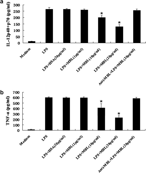
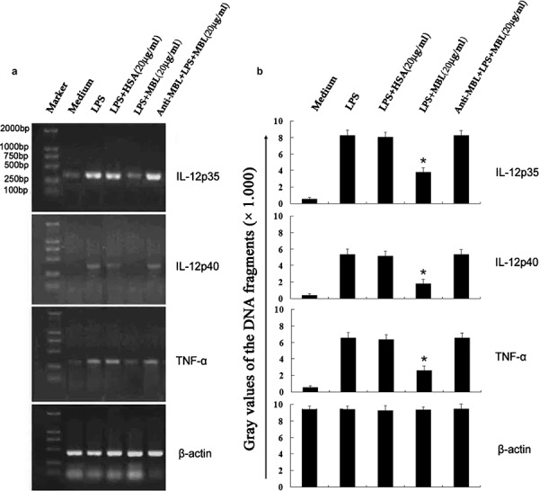
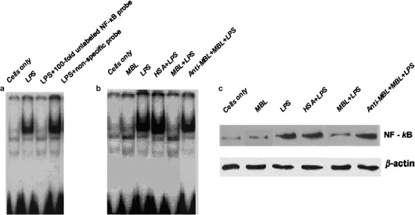
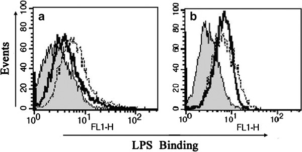
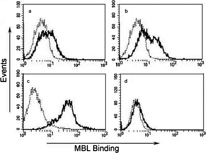
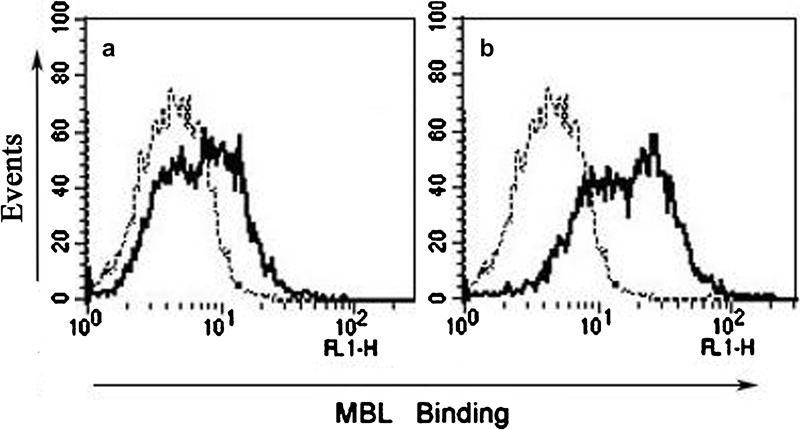
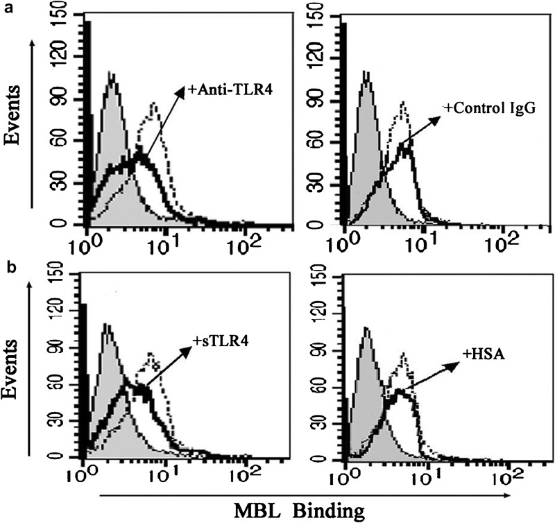
Mannan-binding lectin (MBL) plays a key role in the lectin pathway of complement activation and can influence cytokine expression. Toll-like receptor 4 (TLR4) is expressed extensively and has been demonstrated to be involved in lipopolysaccharide (LPS)-induced signaling. We first sought to determine whether MBL exposure could modulate LPS-induced inflammatory cytokine secretion and nuclear factor-κB (NF-κB) activity by using the monocytoid cell line THP-1. We then investigated the possible mechanisms underlying any observed regulatory effect. Using ELISA and reverse transcriptase polymerase chain reaction (RT-PCR) analysis, we found that at both the protein and mRNA levels, treatment with MBL suppresses LPS-induced tumor-necrosis factor (TNF)-α and IL-12 production in THP-1 cells. An electrophoretic mobility shift assay and western blot analysis revealed that MBL treatment can inhibit LPS-induced NF-κB DNA binding and translocation in THP-1 cells. While the binding of MBL to THP-1 cells was evident at physiological calcium concentrations, this binding occurred optimally in response to supraphysiological calcium concentrations. This binding can be partly inhibited by treatment with either a soluble form of recombinant TLR4 extracellular domain or anti-TLR4 monoclonal antibody (HTA125). Activation of THP-1 cells by LPS treatment resulted in increased MBL binding. We also observed that MBL could directly bind to the extracellular domain of TLR4 in a dose-dependent manner, and this interaction could attenuate the binding of LPS to cell surfaces. Taken together, these data suggest that MBL may affect cytokine expression through modulation of LPS-/TLR-signaling pathways. These findings suggest that MBL may play an important role in both immune regulation and the signaling pathways involved in cytokine networks.


Figures

Figure 1
Figure 1
Analysis of purified MBL protein by SDS–PAGE and WB. (a) Protein marker; (b) purified MBL under non-reducing conditions; (c) purified MBL under reducing conditions; (d) WB analysis of purified MBL under non-reducing conditions; (e) WB analysis of purified MBL under reducing conditions. MBL, mannan-binding lectin; SDS–PAGE, sodium dodecyl sulfate–polyacrylamide gel electrophoresis; WB, western blot.
Figure 2
Figure 2
Inhibition of LPS-induced cytokine production from THP-1 cells by MBL. THP-1 cells were stimulated with LPS (100 ng/ml) in the presence of the indicated concentrations of HSA, anti-MBL pAb and MBL, or MBL alone for 24 h. Supernatants were harvested and subjected to ELISA for IL-12 p40+p70 (a) and TNF-α (b). *P<0.05 as compared to the LPS-stimulated group. Similar results were observed in three independent experiments. HSA, human serum albumin; LPS, lipopolysaccharide; MBL, mannan-binding lectin; pAb, polyclonal antibody; TNF, tumor-necrosis factor.
Figure 3
Figure 3
Inhibition of LPS-induced TNF-α and IL-12 mRNA expression in THP-1 cells by MBL. THP-1 cells were stimulated with LPS (100 ng/ml) in the presence of the indicated concentrations of HSA, anti-MBL pAb and MBL, or MBL alone for 24 h. Samples were taken for RNA extraction from various groups, and the amplification and electrophoresis of TNF-α, IL-12p35 and IL-12p40 genes was carried out simultaneously. (a) Phosphorimage from an individual experiment representing three independent experiments. (b) Gray values of the DNA fragments. *P<0.05 as compared to the LPS-stimulated group. Similar results were observed in three independent experiments. β-actin was used as an internal control. HSA, human serum albumin; LPS, lipopolysaccharide; MBL, mannan-binding lectin; pAb, polyclonal antibody; TNF, tumor-necrosis factor.
Figure 4
Figure 4
Suppression of LPS-stimulated NF-κB activity by MBL. (a) Specific and non-specific competitive assays of NF-κB. THP-1 cells were stimulated with LPS for 1 h, followed by cell harvesting for nuclear extraction. The nuclear extracts were mixed with a radiolabeled NF-κB oligonucleotide probe and 100-fold molar excess of unlabeled consensus NF-κB oligonucleotide and then analyzed by EMSA. A non-specific probe for NF-κB was used as the control. (b) LPS-induced NF-κB DNA-binding activity is inhibited by MBL. THP-1 cells were stimulated with LPS in the presence of 15 µg/ml HSA, anti-MBL pAb and MBL, or MBL for 1 h, followed by cell harvesting to prepare nuclear extracts. The nuclear extracts were mixed with radiolabeled NF-κB oligonucleotide probe and analyzed with EMSA. (c) MBL inhibits NF-κB translocation in THP-1 cells. THP-1 cells were stimulated with LPS in the presence of 15 µg/ml HSA, anti-MBL pAb and MBL, or MBL alone for 1 h, then the cells were harvested to prepare nuclear extracts. The proteins in the nuclei-free supernatants were separated by 10% SDS–PAGE, followed by transfer to a nitrocellulose membrane. After blocking, the membrane was incubated with NF-κB-specific mouse antihuman mAb p65, followed by incubation with HRP-conjugated secondary antibody. ECL was applied for visualization of the protein bands. β-actin was used as an internal control. ECL, enhanced chemiluminescence; EMSA, electrophoretic mobility shift assay; HRP, horseradish peroxidase; HSA, human serum albumin; LPS, lipopolysaccharide; mAb, monoclonal antibody; MBL, mannan-binding lectin; NF, nuclear factor; pAb, polyclonal antibody; SDS–PAGE, sodium dodecyl sulfate–polyacrylamide gel electrophoresis.
Figure 5
Figure 5
Suppression of the binding of smooth LPS to THP-1 cells by MBL. THP-1 cells were pre-incubated with (black solid line) or without (dotted line) MBL for 30 min (a) or with anti-MBL pAb and MBL (black solid line) for 30 min (b) and then further incubated at 4 °C for 30 min with Alexa488-labeled smooth LPS (E. coli O111:B4). The binding of LPS on the cell surface was determined by FCM. The histograms shown are representatives of three experiments. Shaded curves represent the negative control without labeled LPS. FCM, flow cytometry; LPS, lipopolysaccharide; MBL, mannan-binding lectin; pAb, polyclonal antibody.
Figure 6
Figure 6
Ca2+-dependent binding of MBL with THP-1 cells. (a) Tris-buffered saline with 1.3 mM CaCl2; (b) Tris-buffered saline with 5 mM CaCl2; (c) Tris-buffered saline with 10 mM CaCl2; (d) Tris-buffered saline with 5 mM EDTA. Each solution of THP-1 cells was incubated with biotinylated MBL for 10 min before the addition of ExtrAvidin-FITC. After incubation for 30 min at 37 °C, binding of MBL to cells was analyzed by FCM, as shown in the representative histograms (ad) (black lines, biotinylated MBL binding; dotted lines, negative cell-only controls). These data are representative of four independent experiments. FCM, flow cytometry; MBL, mannan-binding lectin.
Figure 7
Figure 7
Enhanced MBL binding after LPS stimulation. THP-1 cells were stimulated for 2 h without (a) or with (b) LPS in complete 1640 medium before addition of biotinylated MBL under conditions of physiological calcium concentrations (1.3 mM). After washing, the cells (2×10⁵) were resuspended. The cell suspension (0.2 ml) was first incubated with biotinylated MBL in Tris-buffered saline containing 1.3 mM CaCl2 for 30 min at room temperature. The cells were then incubated with ExtrAvidin-FITC for 30 min at room temperature. After washing, the cells were analyzed by FCM (black lines, biotinylated MBL binding; dotted lines, negative cell-only controls). These data are representative of four independent experiments. FCM, flow cytometry; LPS, lipopolysaccharide; MBL, mannan-binding lectin.
Figure 8
Figure 8
Enhanced TLR4 expression in THP-1 cells after LPS stimulation. (a) Expression of TLR4 in THP-1 cells was analyzed using RT-PCR. Total RNA was isolated from THP-1 cells cultured in medium (with or without 100 ng/ml LPS) for 2 h using a Qiagen Kit. GAPDH was used as an internal control. (b) Northern blot analysis. Total RNA was prepared from THP-1 cells cultured in medium with or without 100 ng/ml LPS for 2 h. Blots were hybridized with probes specific for TLR4. β-actin was used as an internal control. GAPDH, glyceraldehyde-3-phosphate dehydrogenase; LPS, lipopolysaccharide; RT-PCR, reverse transcriptase polymerase chain reaction; TLR4, Toll-like receptor 4.
Figure 9
Figure 9
Attenuated MBL binding to THP-1 cells by anti-TLR4 mAb or sTLR4. (a) Anti-TLR4 mAb attenuates the binding of MBL to THP-1 cells. THP-1 cells were incubated with anti-TLR4 mAb or mouse isotype IgG (as a control) for 10 min before the addition of biotinylated MBL, and the binding of MBL was detected by ExtrAvidin-FITC (shaded curves, cells only; black solid line, anti-TLR4 mAb or mouse isotype IgG; dotted line, without anti-TLR4 mAb or mouse isotype control IgG). Data are representative of four independent experiments. (b) sTLR4 attenuates the binding of MBL to THP-1 cells. sTLR4 protein or HSA (used as a control) was pre-incubated with biotinylated MBL at 37 °C for 30 min, and the mixture was then incubated with the cells for 30 min on ice before the addition of ExtrAvidin-FITC. Binding of MBL was analyzed by FCM (shaded curves, the cells only; black solid line, sTLR4 or HSA; dotted line, without sTLR4 or HSA). Data shown are representative of four independent experiments. FCM, flow cytometry; HSA, human serum albumin; mAb, monoclonal antibody; MBL, mannan-binding lectin; sTLR4, soluble form of Toll-like receptor 4; TLR4, Toll-like receptor 4.
Figure 10
Figure 10
Binding of MBL to the extracellular domain of TLR4. (a) Ligand blot analysis. Glycosylated and deglycosylated sTLR4 proteins were electrophoresed and transferred onto a PVDF membrane. The proteins were visualized by Coomassie blue staining. The membrane was also incubated with human MBL or HSA, and membrane-bound MBL was detected by anti-MBL mAb, as described in the ‘Materials and methods' section. The oligosaccharide moieties of sTLR4 were visualized using a Carbohydrate Detection Kit (Glycoanalysis). (b) sTLR4 or HSA was coated onto microtiter wells and incubated with the indicated concentrations of MBL. The binding of MBL to sTLR4 was detected by anti-MBL mAb, as described in the ‘Materials and methods' section. The data shown are mean±SE of three experiments. HSA, human serum albumin; mAb, monoclonal antibody; MBL, mannan-binding lectin; PVDF, polyvinylidene difluoride; sTLR4, soluble form of Toll-like receptor 4; TLR4, Toll-like receptor 4.

References

    1. Super M, Thiel S, Lu J, Levinsky RJ, Turner MW. Association of low levels of mannan-binding protein with a common defect of opsonisation. Lancet. 1989;2:1236–1239. - PubMed
    1. Hoffmann JA, Kafatos FC, Janeway CA, Ezekowitz RA. Phylogenetic perspectives in innate immunity. Science. 1999;284:1313–1318. - PubMed
    1. Lu J, Wiedemann H, Timpl R, Reid KB. Similarity in structure between C1q and the collectins as judged by electron microscopy. Behring Inst Mitt. 1993;93:6–16. - PubMed
    1. Palaniyar N, Zhang L, Kuzmenko A, Ikegami M, Wan S, Wu H, et al. The role of pulmonary collectin N-terminal domains in surfactant structure, function, and homeostasis in vivo. . J Biol Chem. 2002;277:26971–26979. - PubMed
    1. Schweinle JE, Nishiyasu M, Ding TQ, Sastry K, Gillies SD, Ezekowitz RA. Truncated forms of mannose-binding protein multimerize and bind to mannose-rich Salmonella montevideo but fail to activate complement in vitro. . J Biol Chem. 1993;268:364–370. - PubMed

Publication types

MeSH terms

LinkOut - more resources