. 2011 Feb 23;6(2):e17397.
doi: 10.1371/journal.pone.0017397.

DHA improves cognition and prevents dysfunction of entorhinal cortex neurons in 3xTg-AD mice

Affiliations

DHA improves cognition and prevents dysfunction of entorhinal cortex neurons in 3xTg-AD mice

Dany Arsenault et al. PLoS One. .

Abstract

Defects in neuronal activity of the entorhinal cortex (EC) are suspected to underlie the symptoms of Alzheimer's disease (AD). Whereas neuroprotective effects of docosahexaenoic acid (DHA) have been described, the effects of DHA on the physiology of EC neurons remain unexplored in animal models of AD. Here, we show that DHA consumption improved object recognition (↑12%), preventing deficits observed in old 3xTg-AD mice (↓12%). Moreover, 3xTg-AD mice displayed seizure-like akinetic episodes, not detected in NonTg littermates and partly prevented by DHA (↓50%). Patch-clamp recording revealed that 3xTg-AD EC neurons displayed (i) loss of cell capacitance (CC), suggesting reduced membrane surface area; (ii) increase of firing rate versus injected current (F-I) curve associated with modified action potentials, and (iii) overactivation of glutamatergic synapses, without changes in synaptophysin levels. DHA consumption increased CC (↑12%) and decreased F-I slopes (↓21%), thereby preventing the opposite alterations observed in 3xTg-AD mice. Our results indicate that cognitive performance and basic physiology of EC neurons depend on DHA intake in a mouse model of AD.


Conflict of interest statement

Competing Interests: The authors have declared that no competing interests exist.

Figures

Figure 1
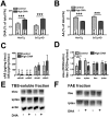
Figure 1. High DHA intake modulates the fatty acid profile of the frontal cortex of NonTg and 3xTg-AD mice and decreases soluble ptau.
High DHA intake from 4 to 14 months of age increased DHA content (A) and decreased AA levels (B). (C) DHA consumption had no effect on the level of Aβ in soluble or insoluble fraction. (D) ptau was decreased in following DHA treatment, without any effect on total tau in soluble fraction and on tau (or ptau) in insoluble fraction. Illustrations of tau/ptau and actin bands in TBS-soluble fraction (E) and tau in FAE fraction (F). Values are expressed as mean ± SEM. Statistical comparisons were performed using two-way ANOVA and correlations were analyzed using a linear regression. Abbreviations: AA, arachidonic acid; DHA, docosahexaenoic acid; FA, fatty acid; FAE, formic-acid extract (insoluble fraction); iAβ, insoluble Aβ; itau, insoluble tau; iptau, insoluble phospho-tau; ROD, relative optical density; sAβ, soluble Aβ; N, number of mice; stau, soluble tau; sptau, soluble phospho-tau; TBS, tris-buffered saline (soluble fraction). *P<0.05, ***P<0.001
Figure 2
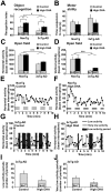
Figure 2. DHA prevents deficits in novel object recognition and episodes of low activity in mice 3xTg-AD.
(A) Horizontal and (B) vertical locomotor activities were both decreased in 3xTg-AD mice compared to NonTg. (C) On the object recognition task, DHA intake improved the cognitive performance of NonTg and 3xTg-AD mice. Recognition index is represented by the percentage of time exploring the object 3 (100*object 3/total exploration during second test). (D) Forelimbs muscle tone necessary to cling onto a stretched cable was comparable between each group, indicating that motor problems did not account for the lower activity of 3xTg-AD mice. (G) 3xTg-AD mice displayed frequent periods of low activity (<15 beam interruptions per 15 sec) or very low activity (<10 beam interruptions per 15 sec), a feature not observed in NonTg mice (H). DHA treatment partly prevented the occurrence of (I) low activity periods and (J) very low activity periods in 3xTg-AD mice. Statistical comparisons were performed using two-way ANOVA or unpaired t-test (low and very low activity periods). Abbreviation: N, number of mice. P<0.05; ***P<0.001
Figure 3
Figure 3. Effects of DHA intake on passive properties of EC deep layer neurons from NonTg and 3xTg-AD mice.
(A) CC of EC neurons increased with DHA intake and decreased with 3xTg-AD expression. The slope of the injected current x time constant versus voltage variation plot was used to estimate CC. Typical examples of slopes are illustrated in panel B. (C) DHA decreased, whereas 3xTg-AD transgenes increased the input resistance following the injection of a hyperpolarized current. Input resistance was determined from the slope of the voltage variation versus injected current plot and examples of current-voltage slope from one cell per group are illustrated in panel D. (E) Membrane conductance was not modulated by diet or transgene expression. (F) Inverse relationship between cell capacitance and ptau in 3xTg-AD mice. (G) DHA intake hyperpolarized EC neurons by altering their resting potential from −60 mV to −70 mV, an effect only present in NonTg mice. (H) Positive relationship between resting potential and TBS-soluble total Aβ (Aβ40 + Aβ42) in 3xTg-AD mice. Values are expressed as mean ± SEM. Statistical comparisons were performed using two-way ANOVA (CC and input resistance) and one-way ANOVA followed by Tukey-Kramer posthoc test (resting potential; variable interaction). Correlation was performed using a linear regression. Recorded neurons were obtained from 8 mice per group. Abbreviations: CC, cell capacitance, n, number of recorded neurons. *P<0.05; ***P<0.001.
Figure 4
Figure 4. Alteration of action potentials characteristics in 3xTg-AD mice: no effect of DHA.
(A) Example of entorhinal cortex (EC) neuron recording following an injection of a 3-s depolarizing current. In this typical trace, the injected current triggered three action potentials. (B) Representation of a post-spike hyperpolarization (zoomed from the dashed square in A). The post-spike hyperpolarization was calculated from the difference between the voltage undershoot after the action potential (dashed line) and the voltage peak of post-spike. (C) Representation of the voltage undershoot following the action potential (zoomed from the dashed square in panel B). Undershoot was the difference between the stabilized voltage after the action potential and the activation threshold. (D) Decreased amplitude was recorded in 3xTg-AD neurons, (E) increased voltage undershoot and (F) reduced post-spike hyperpolarization of EC neurons from 3xTg-AD mice. Values are expressed as mean ± SEM. Statistical comparisons were performed using two-way ANOVA. Recorded neurons were obtained from 8 mice per group. Abbreviation: n, number of recorded neurons. *P<0.05; **P<0.01; ***P<0.001.
Figure 5
Figure 5. DHA treatment and genotype modulate active properties of EC deep layer neurons.
(A, B) Examples of recording trace showing voltage response to a 3-s depolarizing current at the excitation threshold (top trace) and 80±5 pA above the rheobase (bottom trace) from the same neuron of each group. The robust increase in firing frequency detected in 3xTg-AD mice was partly prevented by DHA, which decreased firing frequency in all animals. The relationship between firing rate and injected current (F-I curves) from NonTg or 3xTg-AD neurons were illustrated in the graph at the right of the panel. (C) The steepness of F-I slopes was reduced by DHA intake whereas it was increased by transgene expression. (D) DHA intake increased rheobase only in NonTg mice. Abbreviations: F-I, firing rate versus injected current. Values are expressed as mean ± SEM. Statistical comparisons were performed using two-way ANOVA (F-I curves) and one-way ANOVA followed by Tukey-Kramer posthoc test (rheobase; variable interaction). Recorded neurons were obtained from 8 mice per group. Abbreviation: n, number of recorded neurons. **P<0.01; *** P<0.001.
Figure 6
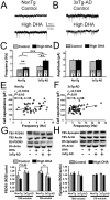
Figure 6. Functional and molecular impairments of glutamatergic synapses in 3xTg-AD mice.
Cellular recordings of (A) NonTg or (B) 3xTg-AD EC neurons fed with the control or high-DHA diet. (C) Transgene expression as well as DHA intake increased the frequencies of sEPSC. (D) The mean amplitude of sEPSC did not differ between groups. CC was significantly correlated with the number of excitatory synaptic events EC neurons from (E) NonTg and (F) 3xTg-AD mice. (G) Postsynaptic protein PSD95 was decreased in detergent-soluble fractions from 3xTg-AD mice whereas DHA had no effect. (H) Diet and transgenes expression did not alter levels of synaptophysin, a presynaptic protein. Numbers of animals per group for molecular experiments were 18-19, except for TBS-soluble synaptophysin (n = 7–8/group). Statistical comparisons were performed using two-way ANOVA. Correlation analyses were performed using linear regressions. Abbreviations: DS, detergent-soluble; TBS, tris-buffered saline; ROD, relative optical density; sEPSC, spontaneous excitatory post-synaptic current. Recorded neurons were obtained from 8 mice per group. Abbreviations: n, number of recorded neurons; N, number of mice. **P<0.01; *** P<0.001.
Figure 7
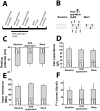
Figure 7. Acute administration of DHA does not replicate the chronic effects of DHA treatment.
(A, B) Timeline and schematic view of DHA application experiments on EC neurons on brain slice. (C, D, E, F) In contrast to chronic oral treatment, acute DHA application on slice had no effect on (C) resting potential, (D) CC, (E) input resistance and, (F) F-I curves. Statistical comparisons were performed using paired t-test. Abbreviations: CC, cell capacitance; F-I, firing rate versus injected current. Recorded neurons were obtained from 5 mice. Abbreviation: n, number of recorded neurons.

References

    1. Duyckaerts C. Looking for the link between plaques and tangles. Neurobiol Aging. 2004;25:735–739; discussion 743-736. - PubMed
    1. Counts SE, Nadeem M, Lad SP, Wuu J, Mufson EJ. Differential expression of synaptic proteins in the frontal and temporal cortex of elderly subjects with mild cognitive impairment. J Neuropathol Exp Neurol. 2006;65:592–601. - PubMed
    1. Scheff SW, Price DA. Alzheimer's disease-related alterations in synaptic density: neocortex and hippocampus. J Alzheimers Dis. 2006;9:101–115. - PubMed
    1. Palop JJ, Chin J, Mucke L. A network dysfunction perspective on neurodegenerative diseases. Nature. 2006;443:768–773. - PubMed
    1. Palop JJ, Chin J, Roberson ED, Wang J, Thwin MT, et al. Aberrant excitatory neuronal activity and compensatory remodeling of inhibitory hippocampal circuits in mouse models of Alzheimer's disease. Neuron. 2007;55:697–711. - PMC - PubMed

Publication types

MeSH terms