

Histologic examination of the eye of acid-sensing ion channel 1a knockout mice
- PMID: 21383900
- PMCID: PMC3047276
Histologic examination of the eye of acid-sensing ion channel 1a knockout mice
Abstract
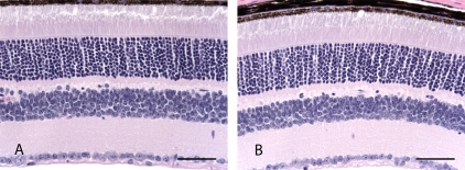
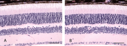
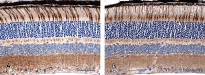
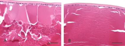
Various subtypes of the acid sensing ion channel have been detected in the retina of rodents and other mammalian species, but the functional importance of this finding is not clearly understood. The purpose of the study was to determine if retinal degeneration was present in ASIC1a-/- mice. The eyes of ASIC1a-/- mice, heterozygote ASIC1a+/- mice, and wild type ASIC1a+/+ mice that were 5 or 22-27 weeks old were processed by routine histotech-nological methods and examined for histologic changes in the retina and other portions of the eye. Additional sections of eyes from ASIC1a-/- and ASIC1a+/+ mice were labeled with peanut agglutinin (PNA) to evaluate cone pho-toreceptors. The retinas of ASIC1a-/-, ASIC1a+/-, and ASIC1a+/+ mice at 5 or 22-27 weeks of age were unremarkable and no morphologic changes in cone photo receptors were detected. Additional findings detected in the eye of ASIC1a+/+ mice included swelling of lens fibers or cataract that were also detected in some of the ASIC1a-/- or ASIC1a+/- mice. Lenticular findings were not considered to be associated with an absence of ASIC1a.
Figures
References
-
- Chang B, Dacey MS, Hawes NL, Hitchcock PF, Milam AH, Atmaca-Sonmez P, Nusinowitz S, Heckenlively JR. Cone photoreceptor function loss-3, a novel mouse model of achromatopsia due to a mutation in Gnat2. Invest Ophthalmol Vis Sci. 2006;47:5017–5021. - PubMed
-
- Ettaiche M, Deval E, Pagnotta S, Lazdunski M, Lingueglia E. Acid-sensing ion channel 3 in retinal function and survival. Invest Ophthalmol Vis Sci. 2009;50:2417–2426. - PubMed
LinkOut - more resources
Full Text Sources
Molecular Biology Databases