. 2011 Feb;7(2):e1001299.
doi: 10.1371/journal.ppat.1001299. Epub 2011 Feb 24.

The C-terminal domain of the Arabinosyltransferase Mycobacterium tuberculosis EmbC is a lectin-like carbohydrate binding module

Affiliations

The C-terminal domain of the Arabinosyltransferase Mycobacterium tuberculosis EmbC is a lectin-like carbohydrate binding module

Luke J Alderwick et al. PLoS Pathog. 2011 Feb.

Abstract

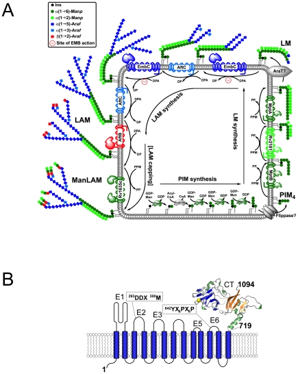
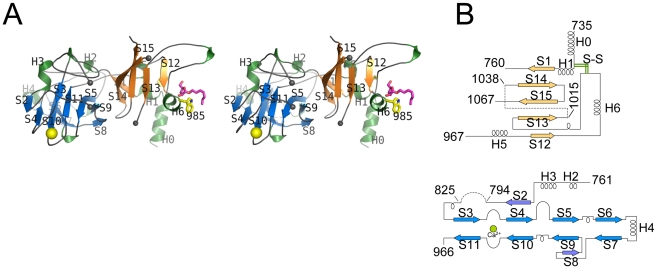
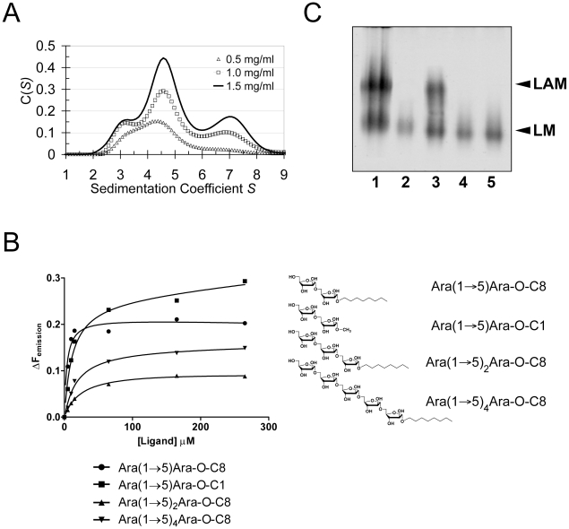
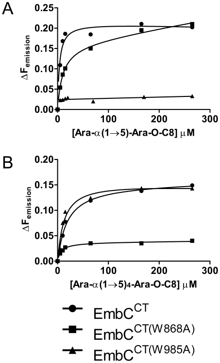
The D-arabinan-containing polymers arabinogalactan (AG) and lipoarabinomannan (LAM) are essential components of the unique cell envelope of the pathogen Mycobacterium tuberculosis. Biosynthesis of AG and LAM involves a series of membrane-embedded arabinofuranosyl (Araf) transferases whose structures are largely uncharacterised, despite the fact that several of them are pharmacological targets of ethambutol, a frontline drug in tuberculosis therapy. Herein, we present the crystal structure of the C-terminal hydrophilic domain of the ethambutol-sensitive Araf transferase M. tuberculosis EmbC, which is essential for LAM synthesis. The structure of the C-terminal domain of EmbC (EmbC(CT)) encompasses two sub-domains of different folds, of which subdomain II shows distinct similarity to lectin-like carbohydrate-binding modules (CBM). Co-crystallisation with a cell wall-derived di-arabinoside acceptor analogue and structural comparison with ligand-bound CBMs suggest that EmbC(CT) contains two separate carbohydrate binding sites, associated with subdomains I and II, respectively. Single-residue substitution of conserved tryptophan residues (Trp868, Trp985) at these respective sites inhibited EmbC-catalysed extension of LAM. The same substitutions differentially abrogated binding of di- and penta-arabinofuranoside acceptor analogues to EmbC(CT), linking the loss of activity to compromised acceptor substrate binding, indicating the presence of two separate carbohydrate binding sites, and demonstrating that subdomain II indeed functions as a carbohydrate-binding module. This work provides the first step towards unravelling the structure and function of a GT-C-type glycosyltransferase that is essential in M. tuberculosis.


Conflict of interest statement

The authors have declared that no competing interests exist.

Figures

Figure 1
Figure 1. Schematic diagram of LAM synthesis and architecture of M. tuberculosis EmbC.
A) Schematic representation of the stepwise assembly of LAM at the membrane of mycobacteria. The precursors of LAM are phosphatidylinositol mannosides (PIM), which contain a phosphatidyl-myo-inositol core unit. Initially, intracellular α-mannosyltransferases catalyse attachment of mannosyl units to inositol, followed by flipping of the glycolipid to the extracellular face of the membrane and further chain extension by membrane-embedded mannosyl- and arabinofuranosyl transferases to generate lipomannan (LM), lipoarabinomannan (LAM) and mannan-capped LAM (ManLAM). Relevant saccharide donor substrates are as follows: GDP-Man (guanosine-5′-diphosphate-α-D-mannose), PPM (C35/C50-polyprenyl-monophospho-mannose), DPA (β-D-arabinofuranosyl-1-monophosphoryl-decaprenol). ManT and AraT designate mannosyl- and arbinosyltransferases that are as yet uncharacterised. B) Topology diagram of EmbC based on the hydropathy analysis with TMHMM (www.cbs.dtu.dk/services/TMHMM/). Extracellular loops are labelled E1-E6 and CT, intracellular loops I1–I7. Functionally important sequence motifs, previously identified in references , , are indicated. The C-terminal domain (residues 719–1094) is shown as a ribbon diagram.
Figure 2
Figure 2. Stereo diagram of EmbCCT and topology of its subdomains.
A) Stereo ribbon diagram of EmbCCT with definition of the secondary structure elements. Grey spheres indicate the boundaries of the disordered loops. The Ca2+ ion (yellow sphere), and positions of Trp985 (yellow sticks) and of the Ara(1→5)Ara-O-C8 ligand (magenta) are shown. B) Topology diagrams of subdomains I (top) and II (bottom), illustrating the connectivity of secondary structure elements and the jelly roll topology of subdomain II.
Figure 3
Figure 3. Metal binding and putative carbohydrate binding sites.
A) Ca2+ site (green sphere) superimposed with an anomalous difference density map (3σ contour level) calculated with in-house diffraction data (CuKα radiation). Metal-ligand interactions are indicated with distances in units of Å. B) σA-weighted Fo−Fc difference density map (3σ contour level) of the Ara(1→5)Ara-O-C8 binding site calculated with phases and calculated amplitudes Fc of the model coordinates prior to incorporation of the ligand. Two symmetry-related molecules are shown (yellow and pink sticks, respectively). Primed residue numbers refer to the symmetry mate. C) Identification of putative carbohydrate binding sites in subdomain II by superimposing EmbCCT with carbohydrate–bound structural homologues. Ligands (shown as stick models) were drawn according to the DALI-alignment with the Cα-traces of structural neighbours. Ligand structures shown in this diagram encompass PDB entries 1ux7, 1w9t, 1o8s, 1w9w, 1uy2, 1od3, 2vzq, 2w47, 2w87, 2cdp, 2cdo, 1uyy, 1uy0, representing the top 10 matches of the DALI search against the PDB90 subset (chains that are less than 90% identical in sequence to each other; Z-scores 6.9–6.3, RMSD 3.0–3.6 Å).
Figure 4
Figure 4. Self-assembly, ligand binding and cell wall analysis.
A) Self-assembly of EmbCCT by analytical ultracentrifugation in sedimentation velocity mode. Protein concentration for the individual distributions is given in units of mg ml−1. Peaks at 3.1S, 4.6S and 7S correspond to fitted molecular weights of 46500 Da, 75800 Da and 138000 Da, respectively. B) Saturation binding of arabinofuranosyl acceptor analogues to EmbCCT probed by intrinsic tryptophan fluorescence. The chemical structures of the ligands are indicated. Data points were fitted to a single site-binding model. C) Effect of substitutions W868A and W985A in full-length M. tuberculosis EmbC on in vivo lipomannan (LM) and LAM synthesis analysed by SDS-PAGE. Lanes are as follows: (1) M. smegmatis wild-type; (2) M. smegmatis ΔembC; (3) M. smegmatis ΔembC+pVV16-Mt-embC; (4) M. smegmatis ΔembC+pVV16-Mt-embCW868A; (5) M. smegmatis ΔembC+pVV16-Mt-embCW985A.
Figure 5
Figure 5. Differential binding of di- and penta-arabinofuranoside acceptor analogues to point mutants of EmbCCT.
Ligand binding was analysed by intrinsic tryptophan fluorescence, comparing saturation binding of wild type EmbCCT, EmbCCT(W868A) and EmbCCT(W985A) for the ligands Ara(1→5)Ara-O-C8 (panel A) and Ara(1→5)4Ara-O-C8 (panel B). Equilibrium dissociation constants derived from non-linear fitting are reported in Table 2.

References

    1. World Health Organisation. 2009. Global Tuberculosis Control: a short update to the 2009 report ( http://www.who.int/entity/tb/publications/2009/factsheet_tb_2009update_d...)
    1. Harries AD, Dye C. Tuberculosis. Ann Trop Med Parasitol. 2006;100:415–431. - PubMed
    1. Jain A, Mondal R. Extensively drug-resistant tuberculosis: current challenges and threats. FEMS Immunol Med Microbiol. 2008;53:145–150. - PubMed
    1. Crick DC, Mahapatra S, Brennan PJ. Biosynthesis of the arabinogalactan-peptidoglycan complex of Mycobacterium tuberculosis. Glycobiology. 2001;11:107R–118R. - PubMed
    1. Briken V, Porcelli SA, Besra GS, Kremer L. Mycobacterial lipoarabinomannan and related lipoglycans: from biogenesis to modulation of the immune response. Mol Microbiol. 2004;53:391–403. - PubMed

Publication types

MeSH terms