. 2011 Feb;7(2):e1001302.
doi: 10.1371/journal.ppat.1001302. Epub 2011 Feb 24.

The bZIP transcription factor MoAP1 mediates the oxidative stress response and is critical for pathogenicity of the rice blast fungus Magnaporthe oryzae

Affiliations

The bZIP transcription factor MoAP1 mediates the oxidative stress response and is critical for pathogenicity of the rice blast fungus Magnaporthe oryzae

Min Guo et al. PLoS Pathog. 2011 Feb.

Erratum in

Abstract

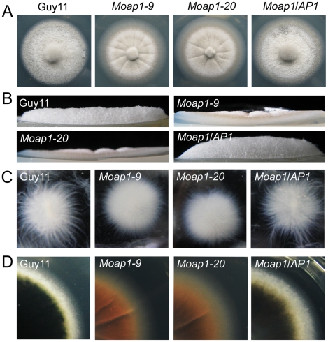
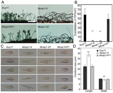
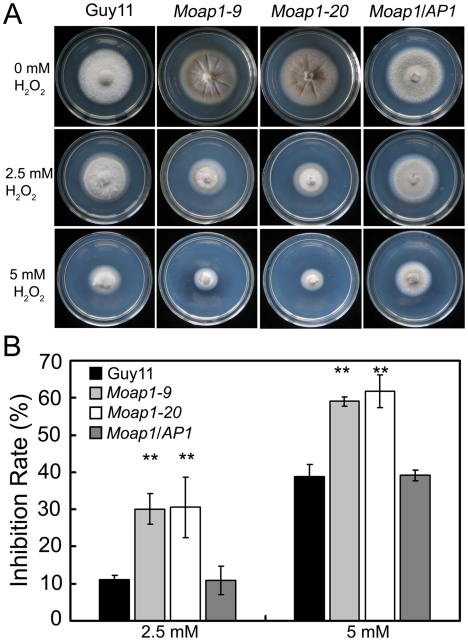
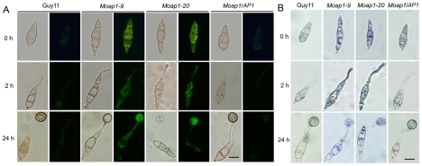
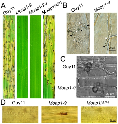
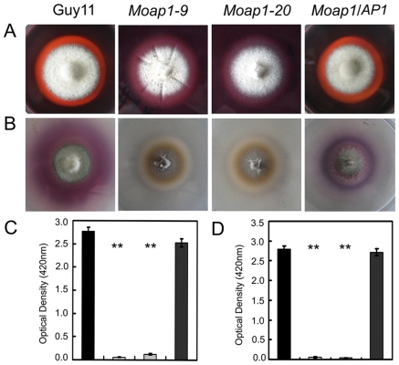
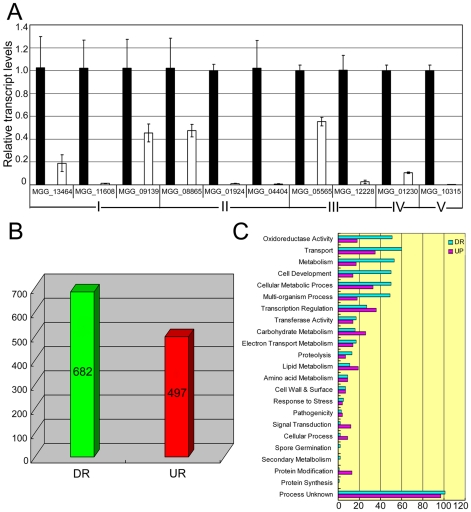
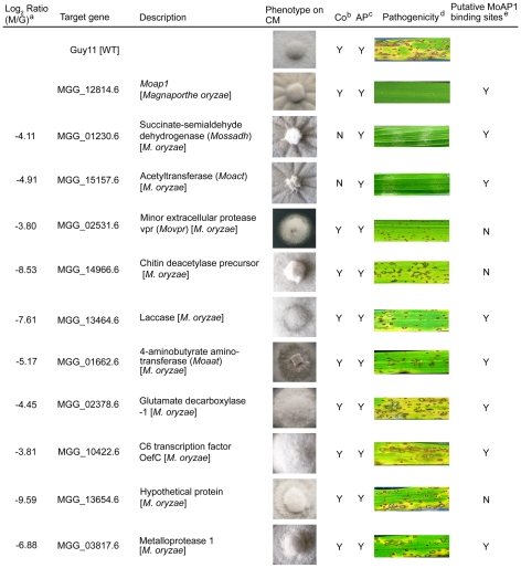
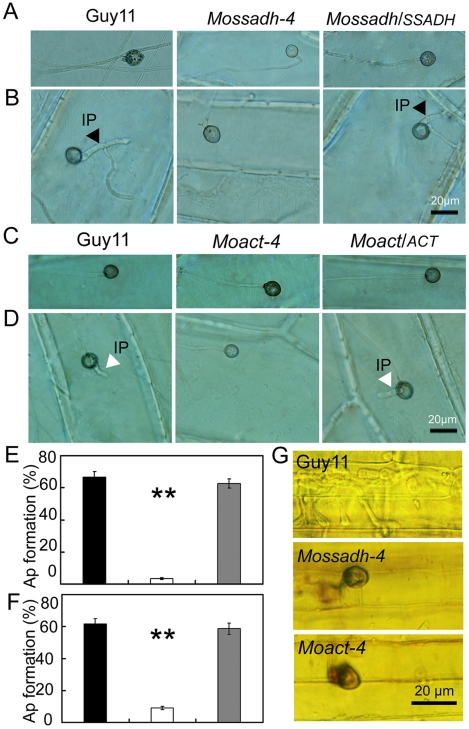
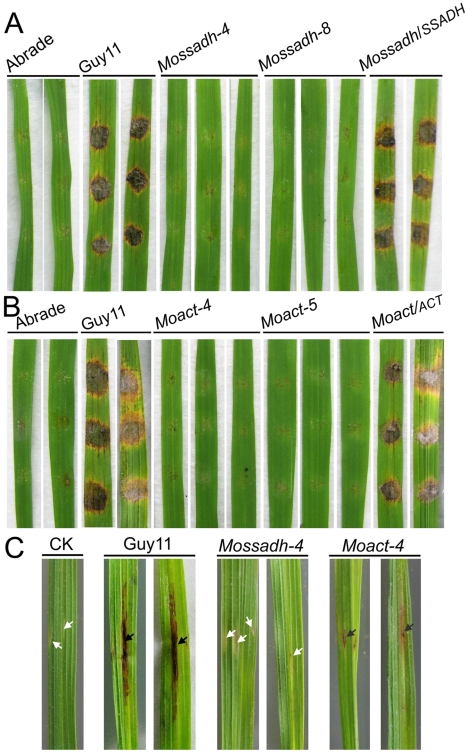
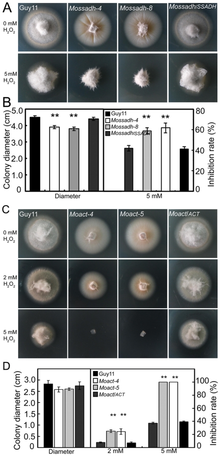
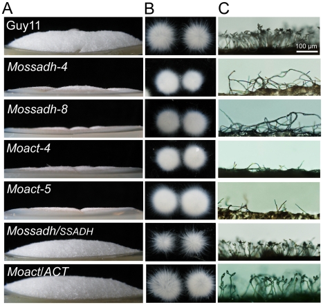
Saccharomyces cerevisiae Yap1 protein is an AP1-like transcription factor involved in the regulation of the oxidative stress response. An ortholog of Yap1, MoAP1, was recently identified from the rice blast fungus Magnaporthe oryzae genome. We found that MoAP1 is highly expressed in conidia and during invasive hyphal growth. The Moap1 mutant was sensitive to H₂O₂, similar to S. cerevisiae yap1 mutants, and MoAP1 complemented Yap1 function in resistance to H₂O₂, albeit partially. The Moap1 mutant also exhibited various defects in aerial hyphal growth, mycelial branching, conidia formation, the production of extracellular peroxidases and laccases, and melanin pigmentation. Consequently, the Moap1 mutant was unable to infect the host plant. The MoAP1-eGFP fusion protein is localized inside the nucleus upon exposure to H₂O₂, suggesting that MoAP1 also functions as a redox sensor. Moreover, through RNA sequence analysis, many MoAP1-regulated genes were identified, including several novel ones that were also involved in pathogenicity. Disruption of respective MGG_01662 (MoAAT) and MGG_02531 (encoding hypothetical protein) genes did not result in any detectable changes in conidial germination and appressorium formation but reduced pathogenicity, whereas the mutant strains of MGG_01230 (MoSSADH) and MGG_15157 (MoACT) showed marketed reductions in aerial hyphal growth, mycelial branching, and loss of conidiation as well as pathogenicity, similar to the Moap1 mutant. Taken together, our studies identify MoAP1 as a positive transcription factor that regulates transcriptions of MGG_01662, MGG_02531, MGG_01230, and MGG_15157 that are important in the growth, development, and pathogenicity of M. oryzae.


Conflict of interest statement

The authors have declared that no competing interests exist.

Figures

Figure 1
Figure 1. The effect of MoAP1 on mycelia growth.
(A) Mycelial growth is not altered in the Moap1 deletion mutant. The Moap1 mutants, the wild type strain (Guy11) and complemented strain (Moap1/AP1) were inoculated on CM medium and cultured at 28°C in darkness for 5 days. (B) Aerial hyphae growth is reduced in the Moap1 mutants. Strains were grown under the same conditions as above and colony side views are displayed. (C) Phenotype of mycelia growth in liquid CM medium. The Moap1 mutants, Guy11, and the complemented strains were inoculated in liquid CM medium for 48 hrs at 28°C in darkness and then photographed. (D). Colony pigmentation is compromised in the Moap1 mutants. The testing strains were cultured as described in Figure 1A and photographed.
Figure 2
Figure 2. MoAP1 disruption leads to abnormal conidial morphology and reduced conidiation.
(A) Development of conidia on conidiophores is affected by Moap1 deletion. Strains grown on RDC medium for 7 days were examined by light microscopy. Bars equal 100 µm. (B) Statistical analysis of conidia production. The conidia produced by the wild type strain (Guy11), the mutants and complemented strains grown on RDC medium for 10 days were collected, counted, and analyzed by Duncan analysis (p<0.01). Asterisks indicate significant differences among Guy11, the Moap1 mutants and complemented strains. Error bar represents standard deviation. (C) Conidium morphology. Conidia were harvested from RDC medium, diluted to 1.0×10⁵ spore/ml, and observed by light microscopy. Bars equal 10 µm. (D) Conidia size comparison. The conidia sizes were determined as width by length from 150 conidia of each strain. Asterisks indicate that the difference is statistically significant. Error bars represent standard deviations.
Figure 3
Figure 3. The Moap1 deletion mutants are hypersensitive to H2O2.
(A) Mycelia growth of the Moap1 mutants under oxidative stress. The wild type strain Guy11, Moap1 mutants and the complemented strain were inoculated on CM medium with or without 2.5 or 5 mM H2O2 and cultured at 28°C for 5 days. (B) The colony diameters of the testing strains were measured and subjected to statistical analysis. The growth inhibition rate is relative to the growth rate of each untreated control [Inhibition rate  =  (the diameter of untreated strain - the diameter of treated strain)/(the diameter of untreated strain ×100%)]. Three repeats were performed and similar results obtained. Error bars represent the standard deviations and asterisks represent significant differences (p<0.01).
Figure 4
Figure 4. The ROS accumulation is compromised in the Moap1 mutant during infection.
(A) Detection of the superoxide by dihydrorhodamine 123 staining. Conidia were inoculated on coverslips and incubated in a moist chamber at 28°C for 0, 2 and 24 hrs before being stained for 2 hrs, rinsed twice with PBS and viewed by epifluorescence microscopy. Fluorescence images were captured using a 100-ms exposure for absorbed light using a GFP filter. Representative bright-field images at each time point are shown. (Scale bars  = 10 µm). (B) Detection of the superoxide by NBT staining. Conidia were prepared as above, stained with a 0.3 mM NBT aqueous solution for 1 hr and viewed by light microscopy. Multiple observations were made and the representative figures were presented (Scale bars  = 10 µm).
Figure 5
Figure 5. Pathogenicity of the Moap1 mutant strain.
(A) The Moap1 deletion mutants lost pathogenicity on rice leaves. 4 ml of conidial suspension (1×10⁵ conidia/ml) for each strain was sprayed on 4-week-old rice seedlings (O. sativa cv CO-39) and 60 healthy rice plants were used in each independent experiment. Diseased leaves were harvested 7 days after inoculation. (B) Onion epidermis cell penetration assay of the Moap1 mutant. The assay was performed by inoculating 30 µl conidia suspension obtained from Guy11, Moap1-9 and the complemented strain. Light microscopic image examination was performed and recorded. Arrows indicate appressoria or invasive hyphae inside cells. (Scale bars  = 20 µm). (C) Rice leaf sheath penetration assay indicating severely confined growth of the Moap1 mutant hyphae at 48 hpi. (Scale bars  = 20 µm). (D) DAB staining indicated the ROS accumulation at the infection site on the rice leaf sheath by the Moap1 mutant at 48 hpi but not by the wild type and complemented strains. (Scale bars  = 20 µm).
Figure 6
Figure 6. Decreased extracellular laccase and peroxidase activities in the Moap1 mutants.
(A) Strains of Guy11, Moap1 mutants and complemented strain were inoculated on CM medium containing Congo Red dye at a final concentration of 200 µg/ml. The discoloration of Congo Red was observed after incubation for 5 days. (B) The laccase activity was monitored in complete media supplemented with 0.2 mM ABTS after 3 days of incubation. (C and D) Guy11, Moap1 mutants and complemented strains were inoculated in CM liquid medium and the laccase activity (C) and the peroxidase activity (D) were measured in the filtrate cultures through ABTS oxidization test with or without H2O2. Dark column indicates Guy11, both white and light grey column equal indicates the Moap1 mutant, and dark gray column indicates the Moap1/AP1 complement strain. Error bars represent the standard deviations and asterisks represent significant differences among the strains tested (p<0.01).
Figure 7
Figure 7. Differential gene expression analysis on transcriptomes of the Moap1 mutant and Guy11 strains.
(A) Real time RT-PCR validated the RNA-SEQ results. Real time RT-PCR was carried out to confirm the RNA-SEQ results through random selection of genes that were down-regulated in the Moap1 mutant. Laccase genes (I), extracellular peroxidase gene (II), and genes involved in the redox homeostasis (III) were down-regulated in the Moap1 mutant. IV and V indicated MGG_01230 (Mossadh) and MGG_10315 (MoMpg1) that were down-regulated. (B) Numbers of altered genes expression in Moap1 mutants. Gene expression profiles were analyzed and 682 genes were down-regulated while 497 genes were upregulated in Moap1 mutants in comparison to the wild type strain (Guy11). Genes whose expression were up or down as indicated by expression profiling were chosen based on the log2 ratio (Moap1/Guy11) values that were either 1.5- fold more or - less. DR and UR denote down- and up-regulation. (C) Functional grouping of genes up- or down-regulated in Moap1 mutants. The up-regulated (in purple) and the down-regulated (in cyan) genes were divided into 23 groups according to their putative functions as described in Materials and Methods.
Figure 8
Figure 8. Phenotype observation and pathogenicity assay of targeted gene deletion mutants.
Ten down-regulated genes in the Moap1 mutant were selected for gene deletion and the phenotype of each mutant was compared and displayed. Gene ID numbers were sourced from www.broad.mit.edu/annotation/genome/magnaporthe_grisea/. a Log2 ratio (Moap1/Guy11) value stands for gene expression fold differences in the Moap1 mutants. b Co represents conidiation and c AP represents appressorium. d The diseased leaves were harvested at 5 dpi, and the results at 7 dpi displayed in Figure S12. e The letter Y stands for yes and N stands for no.
Figure 9
Figure 9. The appressorium-like structure is unable to penetrate the onion epidermis or rice leaf sheath cells.
(A and C) The appressorium-like structure develops at the hyphal tips. The appressorium-like structure was induced by placing the mycelia blocks on the onion epidermis cell for 24 hrs. (Scale bars equal 20 µm). (B and D) Onion epidermis cell penetration assay. The hyphae blocks were inoculated on the onion skin cells for 48 hrs. Penetration was observed using DIC microscopy. (Scale bars equal 20 µm). (E and F) Statistical analysis of appressorium-like structure development. A total of 300 hyphal tips were examined and counted. Dark column indicates Guy11, white column indicates the Mossadh or Moact mutant, and gray column indicates the Mossadh/SSADH or Moact/ACT complement strain. Error bar represents standard deviations and asterisks indicate that the differences were significant. (G) Rice leaf sheath penetration assay. Appressorium like structure formed by the Mossadh mutant and Moact mutant are incapable of penetration into the rice leaf sheath cells 48 hrs post injection (Scale bars  = 20 µm).
Figure 10
Figure 10. Pathogenicity test of Mossadh and Moact mutant strains on the wounded rice leaves.
(A) Pathogenicity test of Mossadh mutants on the wounded rice leaves. The mycelia blocks of the wild type strain Guy11, Mossadh mutants, and the complemented strain were inoculated on the wounded rice leaves and then cultured under moist conditions with 28°C for 5 days. The wounded rice leaves with the CM agar plugs on was used as negative control. This experiment was performed three times with 10 pieces of rice leaves for each strain. Similar results were obtained in each test and this picture showed the representative result. (B) Pathogenicity test of Moact mutants on the wounded rice leaves. The mycelia blocks of Guy11, Moact mutants and the complemented strain were inoculated on the wounded rice leaves and observations made as above. (C) Pathogenicity test of the mutant strain by injection of hyphae fragments. The hyphae fragments of Guy11, the mutant strains and respective complemented strains were prepared as described in Materials and Methods. From left to right, leaves injected with water, wild type strain, Mossadh mutant, and Moact mutant. The arrowheads in black indicate injection sites with necrosis, while the arrowheads in white indicate injection sites without necrosis.
Figure 11
Figure 11. Mossadh and Moact are hypersensitive to H2O2.
(A) Phenotype of the Mossadh mutants under the oxidative stress. The wild type strain, Mossadh mutants and the complemented strain were inoculated on CM agar medium with or without 5 mM H2O2 and cultured at 28°C for 5 days. (B) Statistical analysis of mycelia growth rate with or without H2O2. The analysis was similar to Figure 3B. Error bars represent the standard deviations, and asterisks represent significant differences among Guy11, Mossadh mutants and the complemented strain (p<0.01). (C) Phenotype of the Moact mutant strains under the oxidative stress. Guy11, Moact mutants, and the complemented strain were inoculated on CM agar medium with or without 2 to 5 mM H2O2 and cultured at 28°C for 3 days. (D) Statistical analysis of mycelia growth. Error bars represent the standard deviations, and asterisks indicate that the differences among Guy11, Moact mutants and the complemented strain were statistically significant (p<0.01).
Figure 12
Figure 12. Mossadh and Moact mutants displayed phenotypes similar to the Moap1 mutants.
(A) Aerial hyphae growth reduction in Mossadh and Moact mutants. The wild type, Mossadh mutants, Moact mutants, and respective complemented strains were inoculated on CM medium and cultured at 28°C for 5 days. The aerial hypha was photographed. (B) The phenotype of mycelia grown in liquid CM medium. Strains tested were grown and observed as described in Figure 1C. (C) Development of conidia on conidiophores. Light microscopic observation was performed on strains grown on RDC medium for 7 days. Bars  = 100 µm.

References

    1. Doke N, Miura Y, Sanchez LM, Park HJ, Noritake T, et al. The oxidative burst protects plants against pathogen attack: mechanism and role as an emergency signal for plant bio-defence-a review. Gene. 1996;179:45–51. - PubMed
    1. Apostol I, Heinstein PF, Low PS. Rapid Stimulation of an Oxidative Burst during Elicitation of Cultured Plant Cells: Role in Defense and Signal Transduction. Plant Physiol. 1989;90:109–116. - PMC - PubMed
    1. Chen SX, Schopfer P. Hydroxyl-radical production in physiological reactions. A novel function of peroxidase. Eur J Biochem. 1999;260:726–735. - PubMed
    1. Torres MA, Dangl JL. Functions of the respiratory burst oxidase in biotic interactions, abiotic stress and development. Curr Opin Plant Biol. 2005;8:397–403. - PubMed
    1. Nurnberger T, Brunner F, Kemmerling B, Piater L. Innate immunity in plants and animals: striking similarities and obvious differences. Immunol Rev. 2004;198:249–266. - PubMed

Publication types

MeSH terms