. 2011 Feb 24;6(2):e16408.
doi: 10.1371/journal.pone.0016408.

Clinical impact of down-regulated plasma miR-92a levels in non-Hodgkin's lymphoma

Affiliations

Clinical impact of down-regulated plasma miR-92a levels in non-Hodgkin's lymphoma

Kazuma Ohyashiki et al. PLoS One. .

Abstract

Background: We undertook a study to evaluate the clinical relevance of miR-92a in plasma obtained from non-Hodgkin's lymphoma (NHL) patients, because the miR-17-92 polycistronic miRNA cluster plays a crucial role in lymphomagenesis and affects neo-angiogenesis.

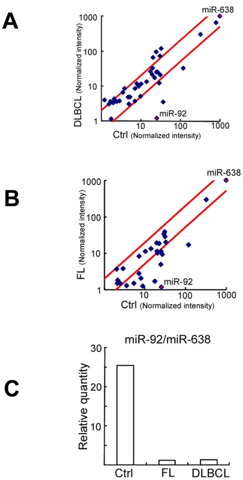
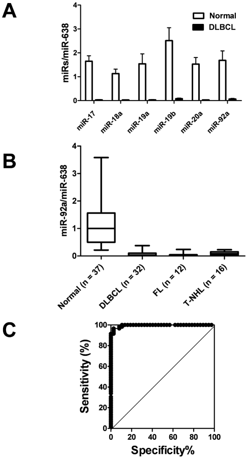
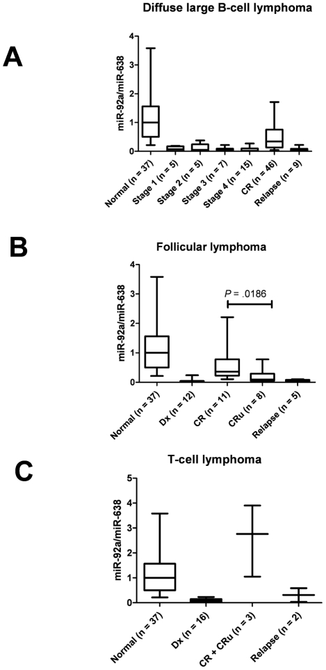
Methodology/principal findings: Plasma miR-92a values in NHL were extremely low (<5%), compared with healthy subjects (P<.0001), irrespective of lymphoma sub-type. The very low plasma level of miR-92a increased in the complete response (CR) phase but did not reach the normal range, and the plasma level was lower again in the relapse phase. Patients in CR or CR unconfirmed with a plasma miR-92a level of less than the cut-off level showed a significantly high relapse rate compared with patients with normalized plasma miR-92a level.

Conclusions/significance: The current results therefore indicate that the plasma miR-92a value could be a novel biomarker not only for diagnosis but also for monitoring lymphoma patients after chemotherapy.


Conflict of interest statement

Competing Interests: The authors have declared that no competing interests exist.

Figures

Figure 1
Figure 1. Comparison of miR expressions in the plasma of normal subjects and non-Hodgkin's lymphoma patients by MiRNA microarray.
Comparison of normalized signal intensities of various miRs in plasmas (miR-638 adjusted to 1000). X axis represents healthy control (Ctrl), and Y axis represents diffuse large B-cell lymphoma (DLBCL) (A) or follicular lymphoma (FL) (B). Red squares show miR-638 (control) and miR-92a. (C) Comparison of the ratio of miR92/miR-638 signal intensity in the plasma of Ctrl, FL, and DLBCL.
Figure 2
Figure 2. Plasma miR-92a expression level and ROC curve in patients with non-Hodgkin's lymphoma by quantitative real-time PCR.
(A) Plasma miR-17-92a expression levels in normal subjects (open bars: Normal: n = 5) and diffuse large B-cell lymphoma (solid bars: DLBCL: n = 13), indicating that all of these miR-17-92a polycistronic miRs were remarkably decreased in plasma obtained from DLBCL patients. Boxes show 95 percentile confidence intervals and lines indicate the range of each miR values. (B) Plasma miR-92a value (miR-92a/miR-638) in healthy subjects, and in patients with diffuse large B-cell lymphoma, follicular lymphoma, and T-cell non-Hodgkin's lymphoma (T-NHL) at diagnosis. Boxes show 95 percentile confidence intervals; lines indicate the range of miR-92a values and also show significantly low levels of plasma miR-92a in various lymphomas, compared with healthy subjects (P<.0001). (C) ROC curve analysis using GraphPad Prism 5.0 software. The cut-off level of plasma miR-92a/miR-638 in all non-Hodgkin's lymphoma patients (n = 60) was 0.2165; the sensitivity was 91.85% (95% CI: 81.32% to 97.19%) and specificity was 97.3% (95% CI: 85.84% to 99.93%).
Figure 3
Figure 3. Plasma miR-92a value (miR-92a/miR-638) in patients with non-Hodgkin's lymphoma at various stages.
(A) Plasma miR-92a value (miR-92a/miR-638) in patients with diffuse large B-cell lymphoma at the time of diagnosis, in the relapse phase, and in the complete remission (CR) state. Decreased plasma miR-92a values recovered in the CR phase (P<.0001) but were still significantly lower than in normal subjects (P<.0001). Plasma miR-92a levels at relapse decreased again and the levels were similar to those at diagnosis. (B) Plasma miR-92a value (miR-92a/638) in patients with follicular lymphoma at the time of diagnosis (Dx), in the relapse phase, and in the CR or complete remission unconfirmed (CRu) states. Patients with CRu status had significantly lower plasma miR-92a/miR-638 values compared with those who had CR (P = .0186 by the Mann-Whitney test). (C) Plasma miR-92a values (miR-92a/miR-638) in patients with T-cell non-Hodgkin's lymphoma at the time of diagnosis, relapse phase, and complete remission (CR and CRu).
Figure 4
Figure 4. In situ MiRNA expression in malignant lymphoma tissues.
(A), (B) and (C) were diffuse large B-cell lymphoma. (D), (E) and (F) were follicular lymphoma. (G), (H) and (I) were normal lymph node. (A), (D), (G), were H&E staining. (B), (C), (E), (F), (H) and (I) were in situ hybridization LNA probes for miR-92a and in the negative control. Blue signals (BCIP/NBT) represent positive results for miR-92a. Nuclear staining (counter stains) was nuclear fast red. We found positive signals for miR-92a in both nucleus and cytoplasm in (B) and (E). We could not detect positive signals in (G) and scrambled control (C), (F) and (I). Bars indicate 50 µm.

References

    1. Baltimore D, Boldin MP, O'Connell RM, Rao DS, Taganov KD. MicroRNAs: new regulators of immune cell development and function. Nat Immunol. 2008;9:839–845. - PubMed
    1. Bartel DP. MicroRNAs: genomics, biogenesis, mechanism, and function. Cell. 2004;116:281–297. - PubMed
    1. He L, Hannon GJ. MicroRNAs: small RNAs with a big role in gene regulation. Nat Rev Genet. 2004;5:522–531. - PubMed
    1. Esquela-Kerscher A, Slack FJ. Oncomirs: microRNAs with a role in cancer. Nat Rev Cancer. 2006;6:259–269. - PubMed
    1. Garzon R, Calin GA, Croce CM. MicroRNAs in cancers. Annu Rev Med. 2009;60:167–179. - PubMed

Publication types

MeSH terms