. 2011 May;20(5):834-48.
doi: 10.1002/pro.609. Epub 2011 Mar 23.

GST pi modulates JNK activity through a direct interaction with JNK substrate, ATF2

Affiliations

GST pi modulates JNK activity through a direct interaction with JNK substrate, ATF2

Anastasia F Thévenin et al. Protein Sci. 2011 May.

Abstract

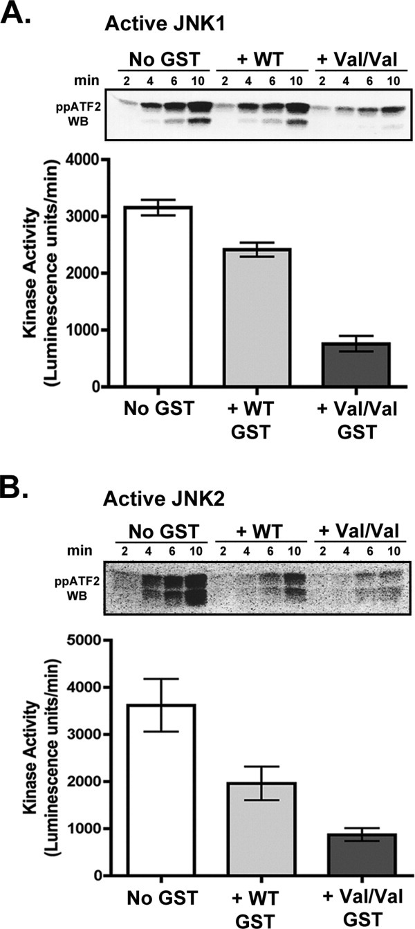
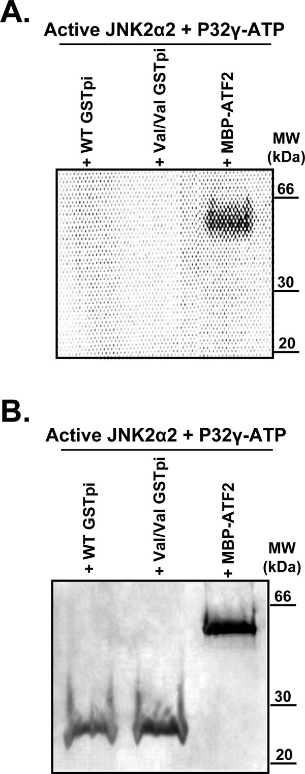
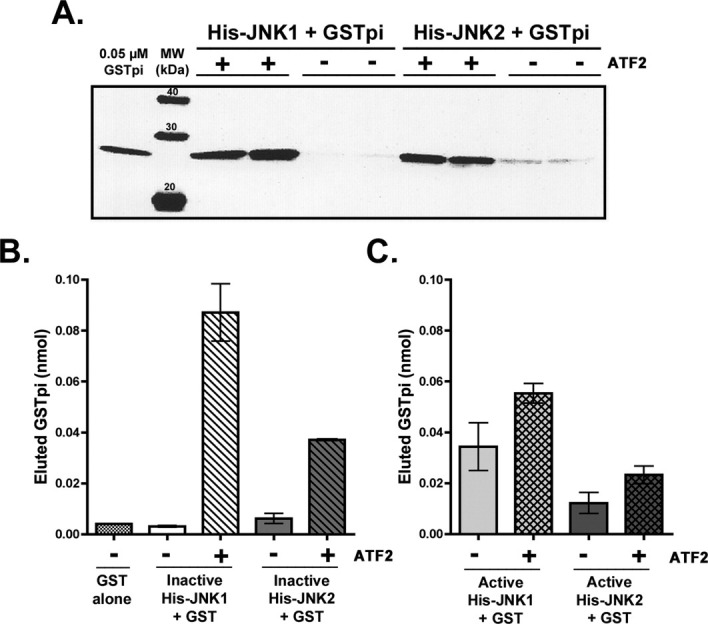
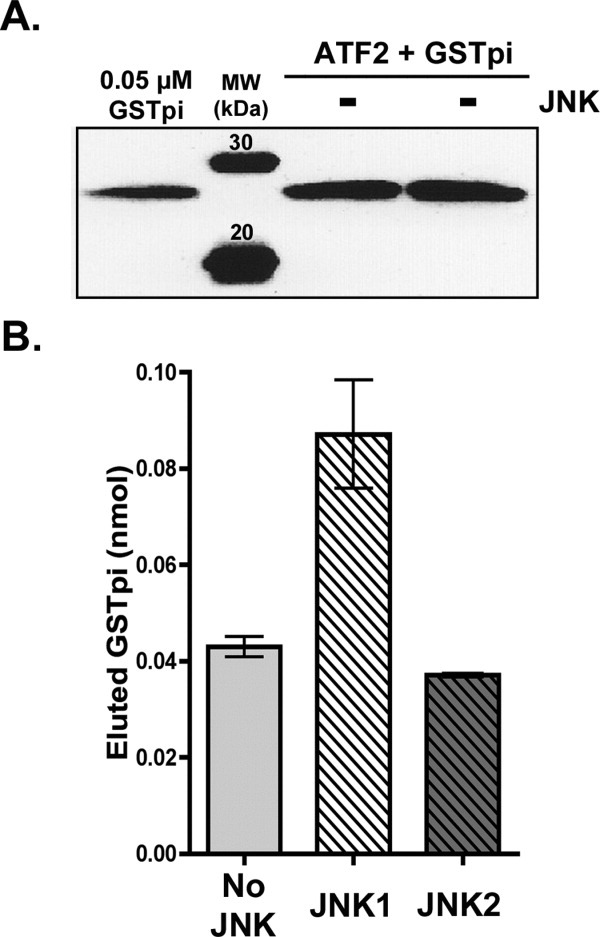
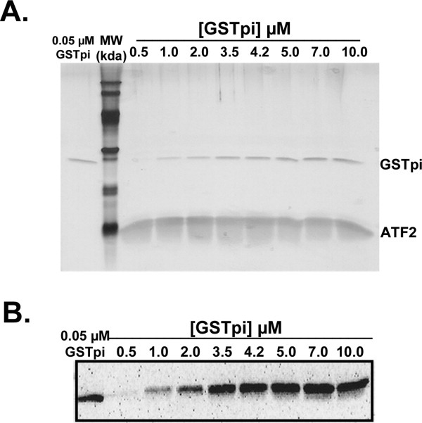
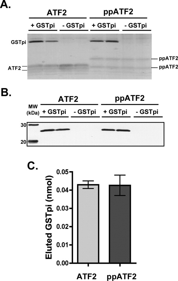
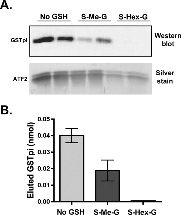
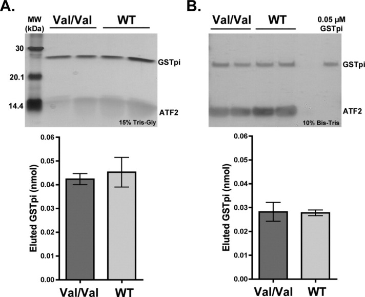
Human GSTpi, an important detoxification enzyme, has been shown to modulate the activity of JNKs by inhibiting apoptosis and by causing cell proliferation and tumor growth. In this work, we describe a detailed analysis of the interaction in vitro between GSTpi and JNK isoforms (both in their inactive and active, phosphorylated forms). The ability of active JNK1 or JNK2 to phosphorylate their substrate, ATF2, is inhibited by two naturally occurring GSTpi haplotypes (Ile105/Ala114, WT or haplotype A, and Val105/Val114, haplotype C). Haplotype C of GSTpi is a more potent inhibitor of JNK activity than haplotype A, yielding 75-80% and 25-45% inhibition, respectively. We show that GSTpi is not a substrate of JNK, as was earlier suggested by others. Through binding studies, we demonstrate that the interaction between GSTpi and phosphorylated, active JNKs is isoform specific, with JNK1 being the preferred isoform. In contrast, GSTpi does not interact with unphosphorylated, inactive JNKs unless a JNK substrate, ATF2, is present. We also demonstrate, for the first time, a direct interaction: between GSTpi and ATF2. GSTpi binds with similar affinity to active JNK + ATF2 and to ATF2 alone. Direct binding experiments between ATF2 and GSTpi, either alone or in the presence of glutathione analogs or phosphorylated ATF2, indicate that the xenobiotic portion of the GSTpi active site and the JNK binding domain of ATF2 are involved in this interaction. Competition between GSTpi and active JNK for the substrate ATF2 may be responsible for the inhibition of JNK catalysis by GSTpi.


Figures

Figure 1
Figure 1
Inhibition of JNK activity by Haplotype A and C GSTpi. Preformed active JNK/ATF2 complexes (in 1:1 molar ratio) were incubated either alone or with 10 μM WT (Haplotype A) or V105/V114 GSTpi (Haplotype C) for 30 min at 25°C. ATP and MgCl2 were added to initiate the JNK catalytic reaction, and the amount of phosphorylation of ATF2 by JNK was monitored as a function of time by Western blot analysis using antibodies against ATF2 phosphorylated at both Thr-69 and Thr-71 (See footnote on page 3). Representative Western blots are shown. Western blot data was analyzed by densitometry in ImageJ to calculate kinase activity in each reaction (in Luminescence units/min). The rates shown in the bar graphs were an average of at least two independent inhibition assays. A, Inhibition of active JNK1. B, Inhibition of active JNK2.
Figure 2
Figure 2
Does JNK catalyze the phosphorylation of GSTpi? To test if active JNK2 was able to phosphorylate GSTpi, incorporation of 32P from [γ32P]ATP into GSTpi was monitored. MBP-ATF2 was used as a positive control since it is known to be phosphorylated by JNK. A, An image generated by the PhosphorImager from a P32 phosphor storage screen. B, Coomassie Blue stained SDS-PAGE gel that was utilized to generate the image in A.
Figure 3
Figure 3
Effect of ATF2 on GSTpi interaction with inactive and active JNKs. Binding experiments were conducted in duplicate, in which either 3.5 μM inactive His-JNK1 or inactive His-JNK2 with or without 3.5 μM untagged ATF2 was used. The binding of V105/V114 GSTpi (7 μM initial) was monitored in the imidazole elutions (as described in the Experimental Procedures). A, Inactive JNKs ± ATF2. Western blotting analysis using antibodies against human GSTpi of the imidazole elutions of the Ni-NTA resin. B, Bar graph of the densitometry results from A, as well as the data for the negative control: GSTpi bound to the Ni-NTA resin lacking any JNK or ATF2. The densitometry value from the GSTpi standard in the Western blot in A (at 0.05 μM), was used (along with volume of the elution) to calculate the amount of GSTpi in the imidazole eluate (in nmol). The total elution volume for samples containing His-JNK1 was 1 mL, while the total elution volume of samples containing His-JNK2 was 0.5 mL. C, Active JNKs ± ATF2. Bar graph of the densitometry analysis of GSTpi binding assays to determine the ability of V105/V114 GSTpi to interact with active JNK1 or active JNK2 in the absence or presence of ATF2. The results are an average of at least two independent binding experiments and were obtained similarly to the results of panel B.
Figure 4
Figure 4
GSTpi binding to active JNK1/ATF2 complexes. Binding experiments in which 3.5 μM ATF2 and 3.5 μM active JNK1 were added to the Ni-NTA resin, and the V105/V114 GSTpi was added in increasing concentrations (from 0.5 to 10 μM). A, Silver staining of the SDS-PAGE gel of the eluted samples of all initial GSTpi concentrations tested. B, Western blot analysis using antibodies against GSTpi of the eluted samples described in panel A.
Figure 5
Figure 5
GSTpi interaction with ATF2 in the absence of JNKs. Resin equilibrated with 3.5 μM ATF2 was mixed with initial 7 μM V105/V114 GSTpi, as was described in the Experimental Procedures. A, The amount of bound GSTpi in the eluted fractions was monitored by Western blotting analysis using antibodies against GSTpi. B, Plots of the densitometry analysis from panel A (no JNK) as well as the results from binding experiments when inactive JNKs were present [from Fig. 3(B)]. The total nmol of eluted GSTpi were calculated as was described for Figure 3.
Figure 6
Figure 6
GSTpi binding to ATF2 with determination of maximum luminescence intensity values. In binding experiments, 3.5 μM ATF2 was added to the Ni-NTA resin, and the V105/V114 GSTpi was added to the resin in increasing initial concentrations (from 0.5 to 10 μM). A. Silver staining of the SDS-PAGE gel of the eluted samples of all initial GSTpi concentrations tested. B. Western blot analysis using antibodies against GSTpi for the eluted samples described in panel A.
Figure 7
Figure 7
Interaction of GSTpi with unphosphorylated and phosphorylated ATF2. Binding experiments between 3.5 μM unphosphorylated or phosphorylated ATF2 (in duplicates) were carried out as was described for previous experiments. A, Silver stained SDS-PAGE gel of the eluted samples from the binding experiment. B, Western blot using GSTpi antibodies of the gel shown in panel A. C, Bar graph of the densitometric analysis of the GSTpi band from panel B to estimate the total nmol of eluted GSTpi.
Figure 8
Figure 8
Effect of S-methyl and S-hexylglutathione on the interaction between GSTpi and ATF2. Binding experiments between GSTpi and ATF2 were carried out either in the regular buffer or in the buffer containing 2.5 mM S-methylglutathione (S-Me-G) or 2.5 mM S-hexylglutathione (S-Hex-G) (as described in the Experimental Procedures). A, Western blot analysis using antibodies against GSTpi of the imidazole eluates. Silver staining was also performed on the gel to demonstrate the corresponding amounts of ATF2 that were found in the imidazole elutions. B, Plot of the densitometry analysis of the Western blot in panel A to show the total eluted GSTpi amounts (nmol).
Figure 9
Figure 9
Comparison of the V105/V114 versus WT GSTpi binding to ATF2. Binding experiments between ATF2 (3.5 μM) and two haplotypes of GSTpi (WT or Haplotype A and V105/V114 or haplotype C) were carried out at two different initial GSTpi concentrations (7 and 2.5 μM). Silver stains of the imidazole elutions, as well as the corresponding plots of the densitometry analysis of GSTpi bands are shown (in nmol of eluted GSTpi). A, 7 μM GSTpi. B, 2.5 μM GSTpi.

References

    1. Holley SL, Fryer AA, Haycock JW, Grubb SE, Strange RC, Hoban PR. Differential effects of glutathione S-transferase pi (GSTP1) haplotypes on cell proliferation and apoptosis. Carcinogenesis. 2007;28:2268–2273. - PubMed
    1. Henderson C, Wolf CR. Disruption of the glutathione transferase Pi class genes. In: Sies H, Packer L, editors. Hoboken, NJ: Methods in enzymology, Vol.401 Elsevier Academic Press; 2005. pp. 116–137. - PubMed
    1. Watson MA, Stewart RK, Smith GB, Massey TE, Bell DA. Human glutathione S-transferase P1 polymorphisms: relationship to lung tissue enzyme activity and population frequency distribution. Carcinogenesis. 1998;19:275–280. - PubMed
    1. Ralat LA, Manevich Y, Fisher AB, Colman RF. Direct evidence for the formation of a complex between 1-cysteine peroxiredoxin and glutathione S-transferase pi with activity changes in both enzymes. Biochemistry. 2006;45:360–372. - PubMed
    1. Townsend D, Findlay V, Tew K. Hoboken, NJ: Methods in enzymology, Vol.401 Elsevier Academic Press; 2005. Glutathione S-transferases as regulators of kinase pathways and anticancer drug targets; pp. 287–307. - PMC - PubMed

Publication types

MeSH terms