. 2011 May;20(5):856-66.
doi: 10.1002/pro.611. Epub 2011 Apr 5.

Altered architecture of substrate binding region defines the unique specificity of UDP-GalNAc 4-epimerases

Affiliations

Altered architecture of substrate binding region defines the unique specificity of UDP-GalNAc 4-epimerases

Veer S Bhatt et al. Protein Sci. 2011 May.

Abstract

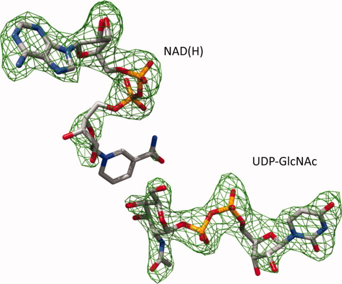
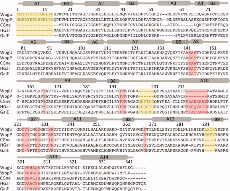
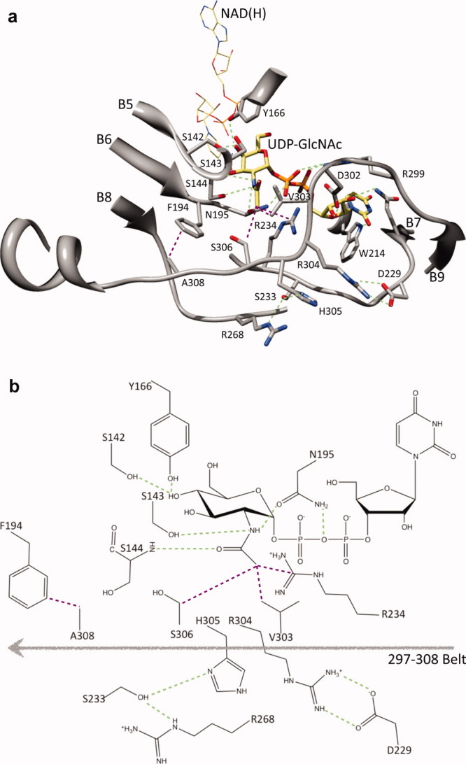
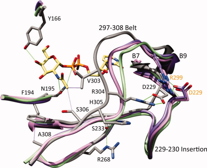
UDP-hexose 4-epimerases play a pivotal role in lipopolysaccharide (LPS) biosynthesis and Leloir pathway. These epimerases are classified into three groups based on whether they recognize nonacetylated UDP-hexoses (Group 1), both N-acetylated and nonacetylated UDP-hexoses (Group 2) or only N-acetylated UDP-hexoses (Group 3). Although the catalysis has been investigated extensively, yet a definitive model rationalizing the substrate specificity of all the three groups on a common platform is largely lacking. In this work, we present the crystal structure of WbgU, a novel UDP-hexose 4-epimerase that belongs to the Group 3. WbgU is involved in biosynthetic pathway of the unusual glycan 2-deoxy-L-altruronic acid that is found in the LPS of the pathogen Pleisomonas shigelloides. A model that defines its substrate specificity is proposed on the basis of the active site architecture. Representatives from all the three groups are then compared to rationalize their substrate specificity. This investigation reveals that the Group 3 active site architecture is markedly different from the "conserved scaffold" of the Group 1 and the Group 2 epimerases and highlights the interactions potentially responsible for the origin of specificity of the Group 3 epimerases toward N-acetylated hexoses. This study provides a platform for further engineering of the UDP-hexose 4-epimerases, leads to a deeper understanding of the LPS biosynthesis and carbohydrate recognition by proteins. It may also have implications in development of novel antibiotics and more economic synthesis of UDP-GalNAc and downstream products such as carbohydrate based vaccines.


Figures

Figure 1
Figure 1
Difference electron density map at the active site of WbgU. Fo-Fc electron density map in the region of UDP-GlcNAc and NAD(H) depicted at an absolute electron density of 0.15 e/Å3 corresponding to 3.0σ cutoff. [Color figure can be viewed in the online issue, which is available at wileyonlinelibrary.com.]
Figure 2
Figure 2
Overall tertiary structure of WbgU/NAD(H)/UDP-GlcNAc complex. N-terminal domain is a modified Rossmann type fold with the cofactor NAD(H) sitting on and parallel to the carboxy edge of the β sheet. The C-terminal domain binds the substrate UDP-GlcNAc. β strands are numbered from the N- to the C- terminal. Dashed grey lines represent the six-residue loop from His287-Ile293 that was not modeled due to poorly defined electron density. [Color figure can be viewed in the online issue, which is available at wileyonlinelibrary.com.]
Figure 3
Figure 3
Multiple sequence alignment between 5 different UDP-hexose 4-epimerases. Regions deemed important for determination of substrate specificity are highlighted in pink. Regions highlighted in yellow are distinct structural variations that do not have a direct bearing on substrate binding or catalysis. WbgU is UDP-GalNAc 4-epimerase from P. shigelloides; WbpP is UDP-GlcNAc 4-epimerase from P. aeruginosa; CGne is UDP-Glc/GlcNAc 4-epimerase (Gne) from C. jejuni; HGal is UDP-Glc/GlcNAc 4-epimerase from Homo sapiens; GalE is UDP-Glc 4-epimerase from E.coli. The numbering and the secondary structure assignment corresponds the structure of WbgU. [Color figure can be viewed in the online issue, which is available at wileyonlinelibrary.com.]
Figure 4
Figure 4
Architecture of the substrate binding region of WbgU. A salt bridge between Arg304 and Asp229 on one side and a hydrogen bonding network formed between His305, Ser233, and Arg268 launch the substrate binding region in a conformation that results in the formation of a hydrophobic cluster contributed by Val303, Arg234, and Ser303. This hydrophobic cluster directly interact with the CH3- group of the GlcNAc moiety. In addition, this conformation results in an interaction between Ala308 and Phe194. This interaction places Asn195 in a hydrogen bond with NH- group of the GlcNAc moiety and the oxy bridge of the diphospho moiety. In addition, a hydrogen bond is formed between Ser143 and NH- group of the GlcNAc moiety and between main chain NH- of Ser144 and CO- group of the GlcNAc moiety. (a) Important interactions with the GlcNAc moiety of the substrate UDP-GlcNAc are highlighted as green (polar interactions) or purple (nonpolar interactions). Tyr166 and Ser142 are hydrogen bonded to 4' hydroxyl group. This region defines the catalytic motif and is highly conserved as is Asn195. The regions defining the substrate binding site are mainly contributed by residues 209-214, 225-234 and 297-308. (b) A schematic representation of the interactions deemed most important in the substrate recognition and catalysis. [Color figure can be viewed in the online issue, which is available at wileyonlinelibrary.com.]
Figure 5
Figure 5
Substrate recognition by the group 1 and the group 2 UDP-hexose 4-epimerases from the perspective of the group 3. His305 in WbgU is substituted by Val in CGne and by Ala in GalE and HGal; Ser233 in WbgU is substituted by Ile in CGne and by Val in GalE and HGal; Arg268 in WbgU is substituted by Gly in CGne, HGal, and GalE (side chains are not shown for clarity). Similarly Arg304 is conserved in WbgU and WbpP whereas being substituted by Ser in CGne, Ala in HGal and Pro in GalE. The loss of the hydrogen bonding network at His305 position and the salt bridge at Arg304 position result in an altered conformation of substrate binding loop in the Group 1 and the Group 2 epimerases. In addition, the insertion of an 8 residue loop at the Asp229 causes formation of a salt bridge with Arg299. The absence of the polar interactions in the Group 1 and the Group 2 epimerases at the 304 and 305 positions in combination with the presence of a salt bridge at the 8 residue insertion between Asp229-Arg299, thus dictates the conformation of the substrate binding region in the group 1 and the group 2 epimerases. The common architecture of the substrate binding region in the group 1 and the group 2 epimerases has one important variation: Ser306 of WbgU (and WbpP) is substituted by a Cys in the Group 2 and by a Tyr in the group 1 hence restricting the access of bulky N-acetyl group to the active site of the group 1 epimerases, which in turn makes them specific towards the nonacetylated substrates. The labels and numbering in black color correspond to WbgU; the labels and numbering in orange color correspond to HGal. [Color figure can be viewed in the online issue, which is available at wileyonlinelibrary.com.]

References

    1. Allard S, Giraud M, Naismith J. Epimerases: structure, function and mechanism. Cell Mol Life Sci. 2001;58:1650–1665. - PMC - PubMed
    1. Bauer AJ, Rayment I, Frey PA, Holden HM. The molecular structure of UDP-galactose 4-epimerase from Escherichia coli determined at 2.5 A resolution. Proteins. 1992;12:372–381. - PubMed
    1. Kowal P, Wang P. New UDP-GlcNAc C4 epimerase involved in the biosynthesis of 2-acetamino-2-deoxy-l-altruronic acid in the O-antigen repeating units of Plesiomonas shigelloides O17. Biochemistry. 2002;41:15410–15414. - PubMed
    1. Ishiyama N, Creuzenet C, Lam JS, Berghuis AM. Crystal structure of WbpP, a genuine UDP-N-acetylglucosamine 4-epimerase from Pseudomonas aeruginosa: substrate specificity in udp-hexose 4-epimerases. J Biol Chem. 2004;279:22635–22642. - PubMed
    1. Holden HM, Rayment I, Thoden JB. Structure and function of enzymes of the Leloir pathway for galactose metabolism. J Biol Chem. 2003;278:43885–43888. - PubMed

Publication types

MeSH terms

Substances

LinkOut - more resources