

Environmentally persistent free radicals decrease cardiac function before and after ischemia/reperfusion injury in vivo
- PMID: 21385100
- PMCID: PMC3152960
- DOI: 10.3109/10799893.2011.555767
Environmentally persistent free radicals decrease cardiac function before and after ischemia/reperfusion injury in vivo
Abstract
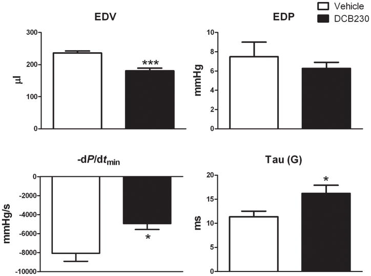
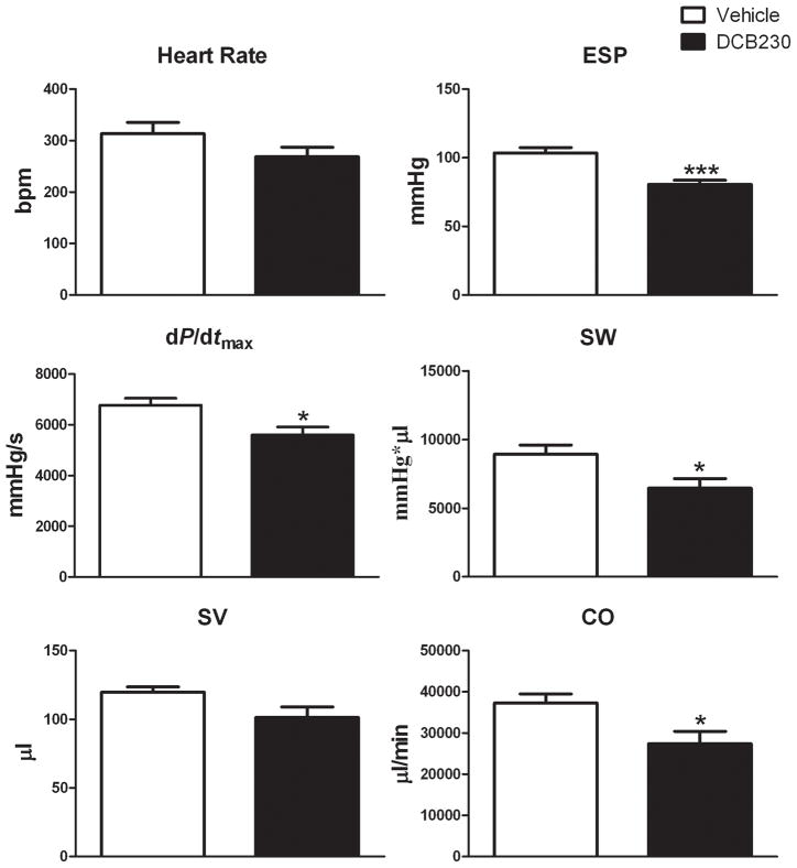
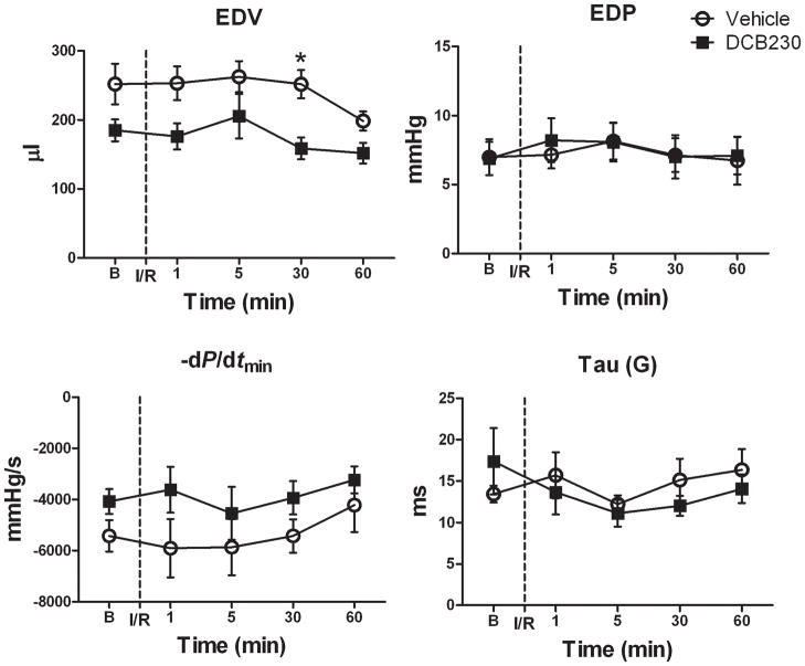
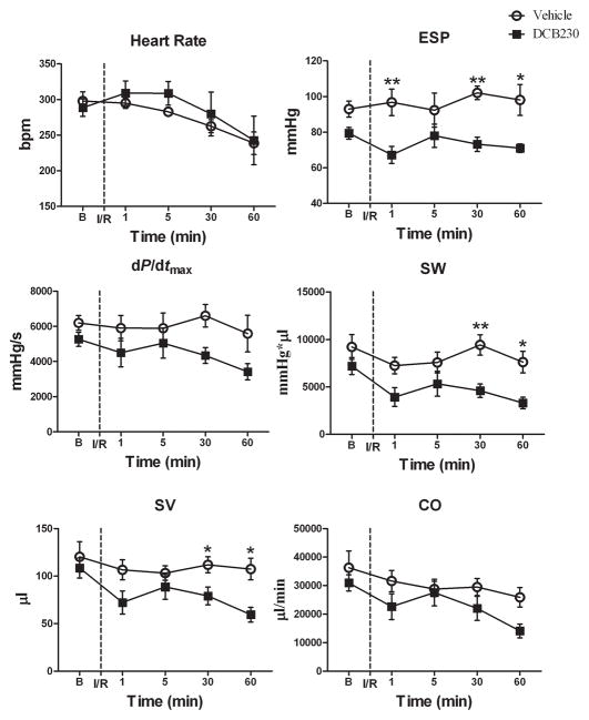
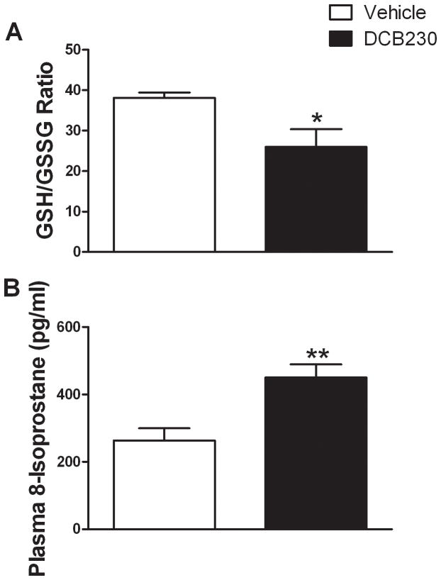
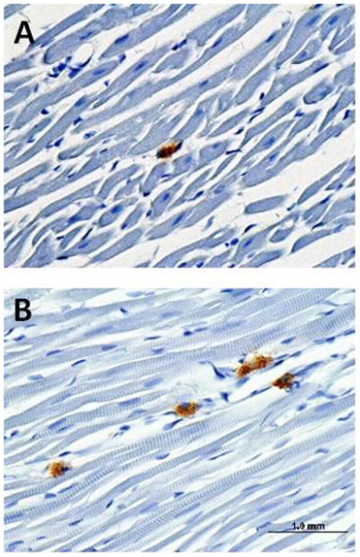
Exposure to airborne particles is associated with increased cardiovascular morbidity and mortality. During the combustion of chlorine-containing hazardous materials and fuels, chlorinated hydrocarbons chemisorb to the surface of transition metal-oxide-containing particles, reduce the metal, and form an organic free radical. These radical-particle systems can survive in the environment for days and are called environmentally persistent free radicals (EPFRs). This study determined whether EPFRs could decrease left ventricular function before and after ischemia and reperfusion (I/R) in vivo. Male Brown-Norway rats were dosed (8 mg/kg, intratracheal) 24 h prior to testing with particles containing the EPFR of 1, 2-dichlorobenzene (DCB230). DCB230 treatment decreased systolic and diastolic function. DCB230 also produced pulmonary and cardiac inflammation. After ischemia, systolic, but not diastolic function was significantly decreased in DCB230-treated rats. Ventricular function was not affected by I/R in control rats. There was greater oxidative stress in the heart and increased 8-isoprostane (biomarker of oxidative stress) in the plasma of treated vs. control rats after I/R. These data demonstrate for the first time that DCB230 can produce inflammation and significantly decrease cardiac function at baseline and after I/R in vivo. Furthermore, these data suggest that EPFRs may be a risk factor for cardiac toxicity in healthy individuals and individuals with ischemic heart disease. Potential mechanisms involving cytokines/chemokines and/or oxidative stress are discussed.
Conflict of interest statement
None declared
Figures
References
-
- Dockery DW, Stone PH. Cardiovascular risks from fine particulate air pollution. N Engl J Med. 2007;356:511–3. - PubMed
-
- Miller KA, Siscovick DS, Sheppard L, Shepherd K, Sullivan JH, Anderson GL, et al. Long-term exposure to air pollution and incidence of cardiovascular events in women. N Engl J Med. 2007;356:447–58. - PubMed
-
- Sun Q, Wang A, Jin X, Natanzon A, Duquaine D, Brook RD, et al. Long-term air pollution exposure and acceleration of atherosclerosis and vascular inflammation in an animal model. JAMA. 2005;294:3003–10. - PubMed
Publication types
MeSH terms
Substances
Grants and funding
LinkOut - more resources
Full Text Sources
Medical