. 2011 Mar 7:9:22.
doi: 10.1186/1741-7015-9-22.

Hypoperfusion of brain parenchyma is associated with the severity of chronic cerebrospinal venous insufficiency in patients with multiple sclerosis: a cross-sectional preliminary report

Affiliations

Hypoperfusion of brain parenchyma is associated with the severity of chronic cerebrospinal venous insufficiency in patients with multiple sclerosis: a cross-sectional preliminary report

Paolo Zamboni et al. BMC Med. .

Abstract

Background: Several studies have reported hypoperfusion of the brain parenchyma in multiple sclerosis (MS) patients. We hypothesized a possible relationship between abnormal perfusion in MS and hampered venous outflow at the extracranial level, a condition possibly associated with MS and known as chronic cerebrospinal venous insufficiency (CCSVI).

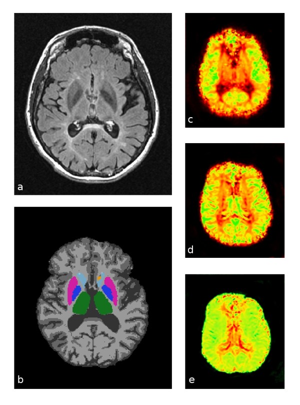
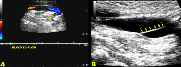
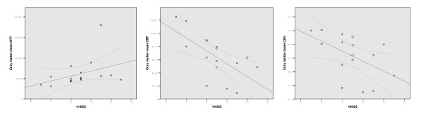
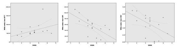
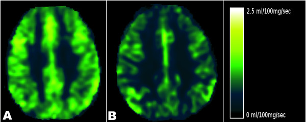
Methods: We investigated the relationship between CCSVI and cerebral perfusion in 16 CCSVI MS patients and 8 age- and sex-matched healthy controls. Subjects were scanned in a 3-T scanner using dynamic susceptibility, contrast-enhanced, perfusion-weighted imaging. Cerebral blood flow (CBF), cerebral blood volume (CBV) and mean transit time (MTT) were measured in the gray matter (GM), white matter (WM) and the subcortical GM (SGM). The severity of CCSVI was assessed according to the venous hemodynamic insufficiency severity score (VHISS) on the basis of the number of venous segments exhibiting flow abnormalities.

Results: There was a significant association between increased VHISS and decreased CBF in the majority of examined regions of the brain parenchyma in MS patients. The most robust correlations were observed for GM and WM (r = -0.70 to -0.71, P < 0.002 and P corrected = 0.022), and for the putamen, thalamus, pulvinar nucleus of thalamus, globus pallidus and hippocampus (r = -0.59 to -0.71, P < 0.01 and P corrected < 0.05). No results for correlation between VHISS and CBV or MTT survived multiple comparison correction.

Conclusions: This pilot study is the first to report a significant relationship between the severity of CCSVI and hypoperfusion in the brain parenchyma. These preliminary findings should be confirmed in a larger cohort of MS patients to ensure that they generalize to the MS population as a whole. Reduced perfusion could contribute to the known mechanisms of virtual hypoxia in degenerated axons.


Figures

Figure 1
Figure 1
Representative source and processed images used for perfusion calculations. (a) Original fluid-attenuated inversion recovery images. (b) Gray, white and deep gray structure segmentations: gray matter in medium gray, white matter in light gray, thalamus in green, globus pallidus in dark blue, putamen in magenta, caudate in light blue and nucleus accumbens in orange. (c) Cerebral blood flow (CBF) map: low flow in red and high flow in green. (d) Cerebral blood volume map: low volume in red and high volume in green. (e) Mean transit time map: short transit time in green and long transit time in red.
Figure 2
Figure 2
Ultrasound assessment in CCSVI. (a) Triplex scanner, longitudinal access of the neck in chronic cerebrospinal venous insufficiency multiple sclerosis patient. In the distal internal jugular vein, close to the junction, the flow is blocked as demonstrated both by the absence of color and by the Doppler spectrum analysis, with the sample completely open in the lumen and no angle correction. (b) An immobile intraluminal defect of the defined septum (multiple arrows) almost completely obstructing the lumen shows the cause of the hampered venous outflow.
Figure 3
Figure 3
Scatterplots showing the relationship between venous hemodynamic insufficiency severity score and gray matter mean transit time (left), cerebral blood flow (center) and cerebral blood volume (right) tissue perfusion parameters in patients with relapsing-remitting multiple sclerosis.
Figure 4
Figure 4
Scatterplots showing the relationship between venous hemodynamic insufficiency severity score and white matter mean transit time (left), cerebral blood flow (center) and cerebral blood volume (right) tissue perfusion parameters in patients with relapsing-remitting multiple sclerosis.
Figure 5
Figure 5
Perfusion MRI study. Left: Cerebral blood flow (CBF) in a 33-year-old, relapsing-remitting (RR) chronic cerebrospinal venous insufficiency (CCSVI) multiple sclerosis (MS) patient with a venous hemodynamic insufficiency severity score (VHISS) of 5. Right: CBF in a 38-year-old, RR CCSVI-MS patient with a VHISS of 12. The dark areas indicate lower CBF in the patient with higher VHISS.

References

    1. Zamboni P, Galeotti R, Menegatti E, Malagoni AM, Gianesini S, Bartolomei I, Mascoli F, Salvi F. A prospective open-label study of endovascular treatment of chronic cerebrospinal venous insufficiency. J Vasc Surg. 2009;50:1348–1358. doi: 10.1016/j.jvs.2009.07.096. e1-e3. - DOI - PubMed
    1. Zamboni P, Galeotti R, Menegatti E, Malagoni AM, Tacconi G, Dall'ara S, Bartolomei I, Salvi F. Chronic cerebrospinal venous insufficiency in patients with multiple sclerosis. J Neurol Neurosurg Psychiatry. 2009;80:392–399. doi: 10.1136/jnnp.2008.157164. - DOI - PMC - PubMed
    1. De Keyser J, Steen C, Mostert JP, Koch MW. Hypoperfusion of the cerebral white matter in multiple sclerosis: possible mechanisms and pathophysiological significance. J Cereb Blood Flow Metab. 2008;28:1645–1651. doi: 10.1038/jcbfm.2008.72. - DOI - PubMed
    1. Varga AW, Johnson G, Babb JS, Herbert J, Grossman RI, Inglese M. White matter hemodynamic abnormalities precede sub-cortical gray matter changes in multiple sclerosis. J Neurol Sci. 2009;282:28–33. doi: 10.1016/j.jns.2008.12.036. - DOI - PMC - PubMed
    1. Trapp BD, Stys PK. Virtual hypoxia and chronic necrosis of demyelinated axons in multiple sclerosis. Lancet Neurol. 2009;8:280–291. doi: 10.1016/S1474-4422(09)70043-2. - DOI - PubMed

Publication types