. 2011 Mar 8;13(2):R25.
doi: 10.1186/bcr2841.

Autoantibodies to aberrantly glycosylated MUC1 in early stage breast cancer are associated with a better prognosis

Affiliations

Autoantibodies to aberrantly glycosylated MUC1 in early stage breast cancer are associated with a better prognosis

Ola Blixt et al. Breast Cancer Res. .

Abstract

Introduction: Detection of serum biomarkers for early diagnosis of breast cancer remains an important goal. Changes in the structure of O-linked glycans occur in all breast cancers resulting in the expression of glycoproteins that are antigenically distinct. Indeed, the serum assay widely used for monitoring disease progression in breast cancer (CA15.3), detects a glycoprotein (MUC1), but elevated levels of the antigen cannot be detected in early stage patients. However, since the immune system acts to amplify the antigenic signal, antibodies can be detected in sera long before the antigen. We have exploited the change in O-glycosylation to measure autoantibody responses to cancer-associated glycoforms of MUC1 in sera from early stage breast cancer patients.

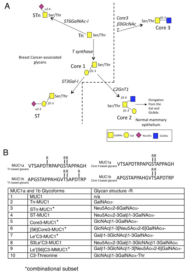
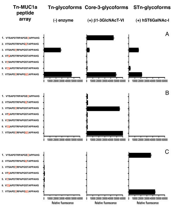
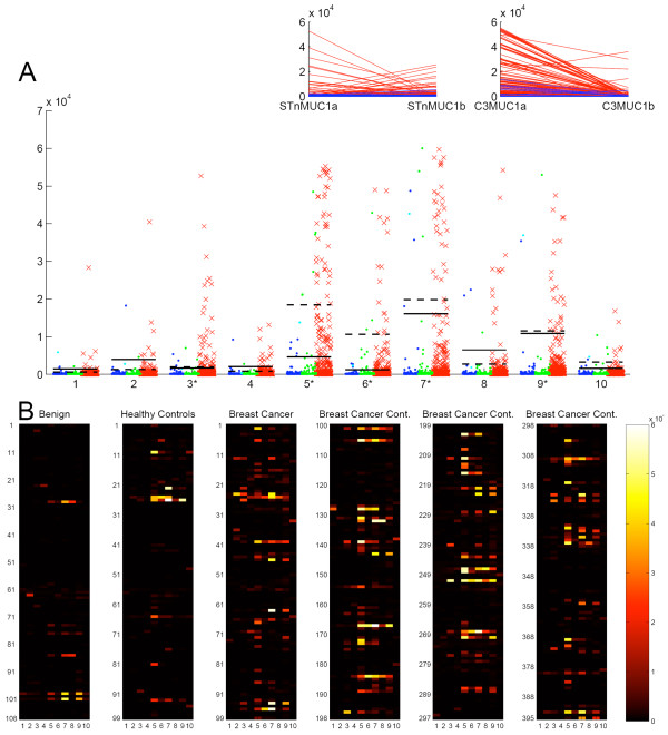
Methods: We used a microarray platform of 60mer MUC1 glycopeptides, to confirm the presence of autoantibodies to cancer associated glycoforms of MUC1 in a proportion of early breast cancer patients (54/198). Five positive sera were selected for detailed definition of the reactive epitopes using on chip glycosylation technology and a panel of glycopeptides based on a single MUC1 tandem repeat carrying specific glycans at specific sites. Based on these results, larger amounts of an extended repertoire of defined MUC1 glycopeptides were synthesised, printed on microarrays, and screened with sera from a large cohort of breast cancer patients (n = 395), patients with benign breast disease (n = 108) and healthy controls (n = 99). All sera were collected in the 1970s and 1980s and complete clinical follow-up of breast cancer patients is available.

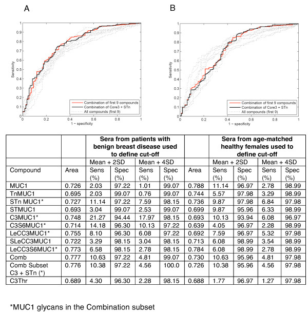
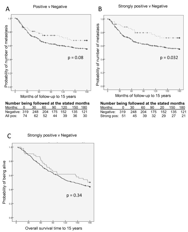
Results: The presence and level of autoantibodies was significantly higher in the sera from cancer patients compared with the controls, and a highly significant correlation with age was observed. High levels of a subset of autoantibodies to the core3MUC1 (GlcNAcβ1-3GalNAc-MUC1) and STnMUC1 (NeuAcα2,6GalNAc-MUC1) glycoforms were significantly associated with reduced incidence and increased time to metastasis.

Conclusions: Autoantibodies to specific cancer associated glycoforms of MUC1 are found more frequently and at higher levels in early stage breast cancer patients than in women with benign breast disease or healthy women. Association of strong antibody response with reduced rate and delay in metastases suggests that autoantibodies can affect disease progression.


Figures

Figure 1
Figure 1
O-linked glycosylation in breast cancer and normal mammary gland and informative glycopeptides used. A, aberrant O-linked glycosylation results in the expression of Tn, STn, Core 1 and ST glycoforms, left of the dotted line. O-glycans in the normal mammary gland are mostly core2 based, right bottom. Note: core3 based glycans (top right) have not been demonstrated in the normal or malignant breast. B, Sequence of MUC1a and MUC1b showing sites of glycosylation and glycans carried.
Figure 2
Figure 2
Epitope mapping of autoantibodies in sera from breast cancer patients. Arrays were produced by on-slide glycosylation of the MUC1a glycopeptides carrying Tn at the sites indicated in red using core3 β3GlcNAc-T6 (middle panel) and ST6GalNAc-I (right panel). The arrays were stained with diluted sera (1:20) and Cy3 labelled anti-human-IgG. A, B, C show data obtained with sera from three individual patients.
Figure 3
Figure 3
Reactivity of auto-antibodies in sera with MUC1 glycoforms. A, dot blot showing the reactivity of auto-antibodies present in sera from breast cancer patients, red crosses; patients with benign breast disease, blue circles; and healthy females aged ranged matched to the cancer patients, green circles. Bars indicate the position for 2x the standard deviation of the mean of the benign serum values, (solid bar) or the aged matched normal values, (dotted bar). Glycopeptides are defined in figure 1B. Inserts show the reactivity of autoantibodies in the sera of breast cancer patients with MUC1a and b carrying STn (left insert) and MUC1a and b carrying core3 (right insert). Each red line represents an individual serum sample. B, heat maps of the reactivity of autoantibodies in sera to glycopeptides 1 to 10 defined in figure 1B. Breast Cancer Cont, breast cancer sera continued.
Figure 4
Figure 4
Receiver operating characteristic analysis of individual and combinations of features. A, sera from patients with benign breast disease defines cut-offs; B, sera from healthy females defines cut-offs. Pale dotted grey lines represent individual features, solid red line represent the combination of all the features and solid black line represents the combinational subset defined in Table by * and Figure 1B by *. The table shows ROC curve area and sensitivity/specificity data for breast cancer sera on each MUC1 glycan, combination of all MUC1 glycans and a combination subset.
Figure 5
Figure 5
Breast cancer patients with autoantibodies to MUC1 glycopeptide combinational subset show increased time to metastasis. A, metastasis free survival with patients with autoantibodies versus no auto-antibodies (P = 0.08). B, metastasis free survival of patients with strong autoantibodies versus no antibodies (P = 0.032). C, overall survival of patients with strong autoantibodies versus no antibodies. Thick lines indicate no auto-antibodies in sera. Dotted lines indicate autoantibodies. Vertical dashes indicate censored events.

References

    1. Desmetz C, Bascoul-Mollevi C, Rochaix P, Lamy PJ, Kramar A, Rouanet P, Maudelonde T, Mangé A, Solassol J. Identification of a new panel of serum autoantibodies associated with the presence of in situ carcinoma of the breast in younger women. Clin Cancer Res. 2009;15:4733–4741. doi: 10.1158/1078-0432.CCR-08-3307. - DOI - PubMed
    1. Hermsen BB, Verheijen RH, Menko FH, Gille JJ, van K, Blankenstein MA, Meijer S, van Diest PJ, Kenemans P, von Mensdorff-Pouilly S. Humoral immune responses to MUC1 in women with a BRCA1 or BRCA2 mutation. Eur J Cancer. 2007;43:1556–1563. doi: 10.1016/j.ejca.2007.04.007. - DOI - PubMed
    1. Chapman C, Murray A, Chakrabarti J, Thorpe A, Woolston C, Sahin U, Barnes A, Robertson J. Autoantibodies in breast cancer: their use as an aid to early diagnosis. Ann Oncol. 2007;18:868–873. doi: 10.1093/annonc/mdm007. - DOI - PubMed
    1. Kufe DW. Functional targeting of the MUC1 oncogene in human cancers. Cancer Biol Ther. 2009;8:1197–1203. - PMC - PubMed
    1. Lumachi F, Basso SM, Brandes AA, Pagano D, Ermani M. Relationship between tumor markers CEA and CA 15-3, TNM staging, estrogen receptor rate and MIB-1 index in patients with pT1-2 breast cancer. Anticancer Res. 2004;24:3221–3224. - PubMed

Publication types