. 2011 Mar 8:9:26.
doi: 10.1186/1479-5876-9-26.

Shipping blood to a central laboratory in multicenter clinical trials: effect of ambient temperature on specimen temperature, and effects of temperature on mononuclear cell yield, viability and immunologic function

Affiliations

Shipping blood to a central laboratory in multicenter clinical trials: effect of ambient temperature on specimen temperature, and effects of temperature on mononuclear cell yield, viability and immunologic function

Walter C Olson et al. J Transl Med. .

Abstract

Background: Clinical trials of immunologic therapies provide opportunities to study the cellular and molecular effects of those therapies and may permit identification of biomarkers of response. When the trials are performed at multiple centers, transport and storage of clinical specimens become important variables that may affect lymphocyte viability and function in blood and tissue specimens. The effect of temperature during storage and shipment of peripheral blood on subsequent processing, recovery, and function of lymphocytes is understudied and represents the focus of this study.

Methods: Peripheral blood samples (n = 285) from patients enrolled in 2 clinical trials of a melanoma vaccine were shipped from clinical centers 250 or 1100 miles to a central laboratory at the sponsoring institution. The yield of peripheral blood mononuclear cells (PBMC) collected before and after cryostorage was correlated with temperatures encountered during shipment. Also, to simulate shipping of whole blood, heparinized blood from healthy donors was collected and stored at 15 °C, 22 °C, 30 °C, or 40 °C, for varied intervals before isolation of PBMC. Specimen integrity was assessed by measures of yield, recovery, viability, and function of isolated lymphocytes. Several packaging systems were also evaluated during simulated shipping for the ability to maintain the internal temperature in adverse temperatures over time.

Results: Blood specimen containers experienced temperatures during shipment ranging from -1 to 35 °C. Exposure to temperatures above room temperature (22 °C) resulted in greater yields of PBMC. Reduced cell recovery following cryo-preservation as well as decreased viability and immune function were observed in specimens exposed to 15 °C or 40 °C for greater than 8 hours when compared to storage at 22 °C. There was a trend toward improved preservation of blood specimen integrity stored at 30 °C prior to processing for all time points tested. Internal temperatures of blood shipping containers were maintained longer in an acceptable range when warm packs were included.

Conclusions: Blood packages shipped overnight by commercial carrier may encounter extreme seasonal temperatures. Therefore, considerations in the design of shipping containers should include protecting against extreme ambient temperature deviations and maintaining specimen temperature above 22 °C or preferably near 30 °C.


Figures
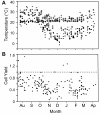
Figure 1
Figure 1
Recorded internal package temperatures during shipment and cell yields of blood from off-site cancer centers. (A) High (+) and low (●) package temperatures recorded between August, 2005 through April, 2006. (B) Yield of PBMC (cell yield) obtained from specimens shipped during this time after Ficoll separation. The ratio cell yield is expressed as a ratio of total number of PBMC collected after Ficoll divided by the number of PBMC (lymphocytes and monocytes) estimated from the differential WBC recorded on the same specimens before shipment. The dashed line represents 100% recovery of PBMC after Ficoll as a ratio cell yield of one.
Figure 2
Figure 2
The recovery of cells after Ficoll separation increased as shipping temperature increased. (A) Correlation of the ratio cell yield with minimum temperature during transport; p = 0.001. (B) Correlation of the ratio cell yield with maximum temperature during transport; p = 0.04. (C) Correlation of the ratio cell yield as a function of maximum temperature deviation from room temperature (22°C) during shipment; p < 0.001
Figure 3
Figure 3
Viability of PBMC 24 hours after thawing from liquid nitrogen. After whole blood was incubated at different temperatures for varying lengths of time, PBMC were isolated and cryopreserved. Samples were thawed and rested overnight at 37°C before staining with CD4, CD8, Annexin V and 7-AAD. The viable populations were defined as Annexin V negative and 7AAD negative and are expressed as a percentage of the respective populations of (A) PBMC, (B) CD8 and (C) CD4 lymphocytes. Shaded area on graph represents the control condition of incubating whole blood at 22°C for 24 hours to which all other conditions were compared. (*) p = 0.003; (**) p = 0.03.
Figure 4
Figure 4
CD8 T cells show greater susceptibility to apoptosis than CD4 T cells. The percentage of cells in different stages of apoptosis was evaluated for CD4 and CD8 T cell populations. (A) Percentage of CD4 lymphocytes in early stages of apoptosis (Annexin V+, 7AAD-) and (B) late stages of apoptosis (Annexin V+, 7AAD+); (C) CD8 lymphocytes in early stages of apoptosis (Annexin V+, 7AAD-) and (D) late stages of apoptosis (Annexin V+, 7AAD+). Shaded region indicates control condition as described in Figure 3.
Figure 5
Figure 5
Mitogen and antigen-activated PBMC responses as detected by IFNgamma secretion in an ELIspot assay. After thawing from liquid nitrogen, PBMC were incubated 18 hours at 37°C with (A) PMA/ionomycin, (B) PHA or (C) CEF peptide pool and then tested for IFNg secretion by ELIspot assay. Results are presented as SFC per 200,000 PBMC for PMA and PHA. CEF SFC are adjusted for the percentage of CD8+ T cells and presented as SFC per 200,000 CD8 T cells. Each condition is compared to the control condition (arrows) as described in Figure 3. (*) p < 0.004.
Figure 6
Figure 6
Internal temperature change over time in containers designed for shipping blood specimens. Ten water-filled vacutainer vials were pre-warmed to 37°C placed inside the JVI Control Temp shipping container or in the Safeguard (SG) shipping container, surrounded with pre-warmed gel packs, placed inside an insulated corrugated cardboard container, and sealed with tape for testing at low external temperatures. Internal package temperatures were continuously monitored inside JVI and SG shipping containers while placed (A) at a constant low temperature of 4°C for 22 hours followed by 22°C for 8 hours; (B) outdoors in ambient winter temperatures for 16 hours; and (C) outdoors in ambient winter temperatures for 18 hours followed by placement of package at 22°C for 20 hours. (green diamond) External package (ambient) temperature; internal package temperatures: (red triangle), JVI with 37°C thermal pack; (purple square), SG with 37°C thermal pack; (blue triangle), JVI with 22°C thermal pack; (blue square), SG with 22°C thermal pack. Solid black line indicates 15°C; dashed line denotes 0°C.
Figure 7
Figure 7
Temperature performance test of the JVI Control Temp shipping container. Five vials, filled with water conditioned at 20°C, were suspended inside the foam vial holder and placed inside the plastic clamshell plastic box fitted with small foam pads. Two of the vials each had a T thermocouple taped to it. The clamshell package was put inside the insulated corrugated cardboard box in which two 12 oz. Control Temp gel packs conditioned at 20°C were also placed inside and taped shut. The shipping container was set inside a 45°C chamber for forty-five hours and the internal package temperature recorded as described in Methods. The red line indicates the external temperature of the chamber. The blue line represents the average internal temperature of the shipping container obtained from duplicate temperature probes.

References

    1. Vaught JB. Blood collection, shipment, processing, and storage. Cancer Epidemiol Biomarkers Prev. 2006;15:1582–1584. doi: 10.1158/1055-9965.EPI-06-0630. - DOI - PubMed
    1. Vaught JB, Caboux E, Hainaut P. International efforts to develop biospecimen best practices. Cancer Epidemiology Biomarkers & Prevention. 2010;19:912–915. - PubMed
    1. Hallmans G, Vaught JB. In: Methods in Biobanking. Dillner J, editor. Springer Science+Business Media, LLC; 2011. Best Practices for Establishing a Biobank; pp. 241–260. full_text. - PubMed
    1. Leyland-Jones BR, Ambrosone CB, Bartlett J, Ellis MJC, Enos RA, Raji A, Pins MR, Zujewski JA, Hewitt SM, Forbes JF, Abramovitz M, Braga S, Cardoso F, Harbeck N, Denkert C, Jewell SD. Recommendations for collection and handling of specimens from group breast cancer clinical trials. J Clin Oncol. 2008;26:5638–5644. doi: 10.1200/JCO.2007.15.1712. - DOI - PMC - PubMed
    1. Ashmore LM, Shopp GM, Edwards BS. Lymphocyte subset analysis by flow cytometry. Comparison of three different staining techniques and effects of blood storage. Journal of Immunological Methods. 1989;118:209–215. doi: 10.1016/0022-1759(89)90008-2. - DOI - PubMed

Publication types