. 2011 May 15;353(2):266-74.
doi: 10.1016/j.ydbio.2011.02.029. Epub 2011 Mar 6.

Hox genes define distinct progenitor sub-domains within the second heart field

Affiliations

Hox genes define distinct progenitor sub-domains within the second heart field

Nicolas Bertrand et al. Dev Biol. .

Abstract

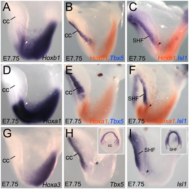
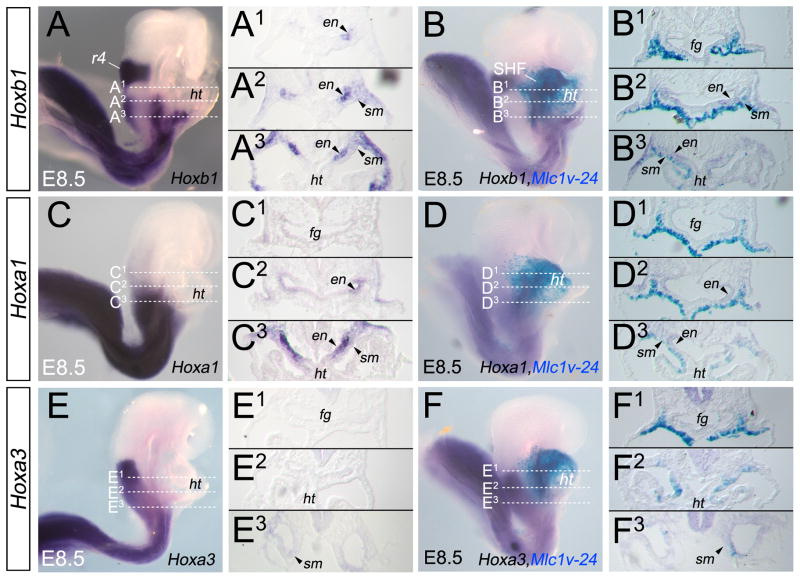
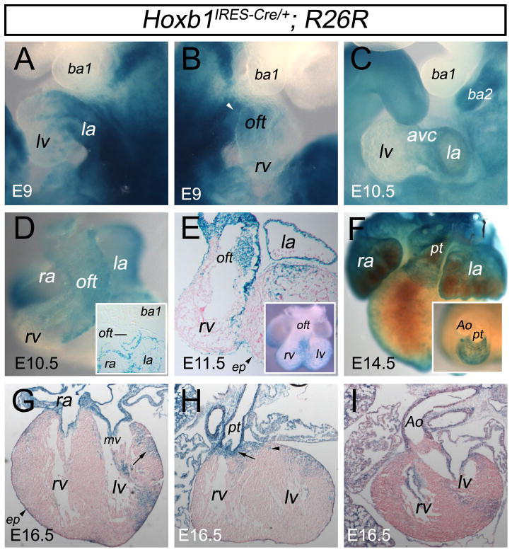
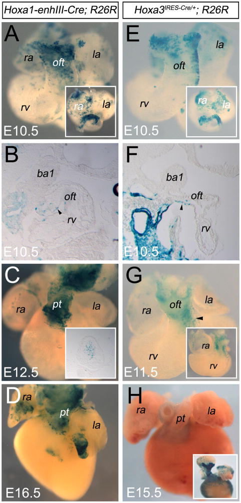
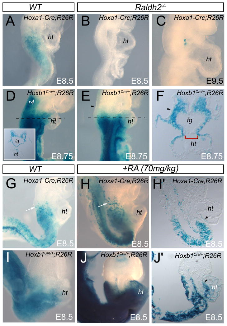
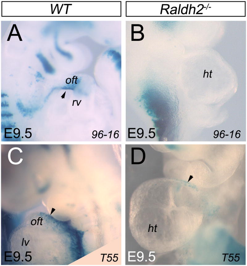
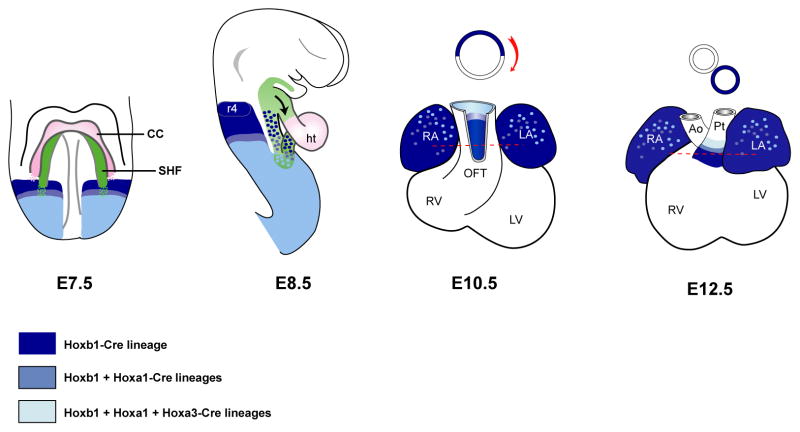
Much of the heart, including the atria, right ventricle and outflow tract (OFT) is derived from a progenitor cell population termed the second heart field (SHF) that contributes progressively to the embryonic heart during cardiac looping. Several studies have revealed anterior-posterior patterning of the SHF, since the anterior region (anterior heart field) contributes to right ventricular and OFT myocardium whereas the posterior region gives rise to the atria. We have previously shown that Retinoic Acid (RA) signal participates to this patterning. We now show that Hoxb1, Hoxa1, and Hoxa3, as downstream RA targets, are expressed in distinct sub-domains within the SHF. Our genetic lineage tracing analysis revealed that Hoxb1, Hoxa1 and Hoxa3-expressing cardiac progenitor cells contribute to both atria and the inferior wall of the OFT, which subsequently gives rise to myocardium at the base of pulmonary trunk. By contrast to Hoxb1(Cre), the contribution of Hoxa1-enhIII-Cre and Hoxa3(Cre)-labeled cells is restricted to the distal regions of the OFT suggesting that proximo-distal patterning of the OFT is related to SHF sub-domains characterized by combinatorial Hox genes expression. Manipulation of RA signaling pathways showed that RA is required for the correct deployment of Hox-expressing SHF cells. This report provides new insights into the regulatory gene network in SHF cells contributing to the atria and sub-pulmonary myocardium.


Figures

Figure 1
Figure 1
Relation between Hoxb1 and Hoxa1 expression and the heart fields. (A–F) Hoxb1 and Hoxa1 expression analysis by single and double in situ hybridizations (ISH) on E7.75 embryos. (G) Hoxa3 expression analysis by ISH. (H,I) Whole-mount ISH with Tbx5 and Islet1 (Isl1) probes, which mark the cardiac crescent (cc) and the second heart field (SHF) respectively. Insets display a ventral view of same stained embryo. (A,D,G) At E7.75, Hoxb1, Hoxa1 and Hoxa3 reach their most anterior border of expression near the cardiac crescent (cc). (B,C) Whole-mount ISH analysis showing that the anterior border of Hoxb1 expression overlaps with Isl1 (arrowhead in C), but not with Tbx5. (E,F) Whole-mount ISH analysis showing Hoxa1 expression in an adjacent domain of Tbx5 and Isl1 (arrowhead in F). (G) Anterior border of Hoxa3 expression is posterior to Tbx5 and Isl1 regions. cc, cardiac crescent; SHF, second heart field.
Figure 2
Figure 2
Hoxb1 and Hoxa1 expression patterns define distinct sub-domains within the second heart field. (A–F) Lateral views of embryos at E8.5. (A,C,E) Whole-mount in situ hybridization (ISH) analysis of Hoxb1 (A), Hoxa1 (C) and Hoxa3 (E) mRNAs. (B,D,F) Whole-mount ISH analysis of Hoxb1 (B), Hoxa1 (D) and Hoxa3 (F) genes combined with X-gal staining for the Mlc1v-nlacZ-24 transgene, which marks the anterior heart field (AHF). Dotted lines in A–F indicate the plane of sections in A1–F2. (A1–A3) Sections showing expression of Hoxb1 in the medial (A2, arrowhead) and posterior (A3) domains of the second heart field (SHF), and the absence of expression in the anterior domain of the SHF (A1). Note the expression of Hoxb1 in the anterior foregut endoderm. (B1–B3) Sections showing co-localization of Hoxb1 and X-gal staining in the caudal region of the AHF (B2,B3) but not in the anterior region of the AHF (B1). (C1–C3) Expression of Hoxa1 is only detected in the posterior region of the SHF (C3, arrowheads). Note the expression of Hoxa1 in the anterior foregut endoderm (C2, C1). (D1–D3) Sections showing the co-localization of Hoxa1 and X-gal labeled cells only in the posterior region of the AHF (D3, arrowhead). (E1–E3) Hoxa3 expression is observed in the splanchnic mesoderm (E3, arrowhead) located posteriorly to the heart tube. (F1–F3) Sections showing that Hoxa3 is not detected in the AHF. cc, cardiac crescent; en, endoderm; fg, foregut; ht, heart tube; SHF, second heart field; sm, splanchnic mesoderm.
Figure 3
Figure 3
Genetic lineage analysis reveals a contribution of Hoxb1+ cardiac progenitors to the atria and the myocardium at the base of the pulmonary trunk. (A–I) Hoxb1-lineage visualized by X-gal staining of Hoxb1IRES-Cre; R26R-lacZ embryos. (A–C) Lateral views of X-gal stained embryos at E9 (A,B) and E10.5 (C). (D–F) Ventral views of X-gal stained hearts at E10.5 (D), E11.5 (E) and E14.5 (F). (G,I) Transverse sections of X-gal stained hearts at E16.5. (A,B) X-gal staining showing a contribution of Hoxb1-positive cells to the venous pole (left atrium) and arterial pole (white arrowhead) of Hoxb1IRES-Cre; R26R-lacZ hearts. (C) Lateral view of an E10.5 X-gal stained embryo, showing β-galactosidase activity in the left atrium and the atrioventricular canal. Note that neural crest derivatives populate the second branchial arch (ba2). (D) Ventral view of X-gal stained heart from Hoxb1IRES-Cre; R26R-lacZ embryo at E10.5. β-galactosidase activity is detected in the left and right atria but also in the outflow tract (OFT). Inset is a frontal section through stage E10.5 embryo, showing β-galactosidase activity only in the inferior wall of the OFT. (E) Transverse section of the heart from an E11.5 Hoxb1IRES-Cre; R26R-lacZ embryo. β-galactosidase activity is detected in the atria, the epicardium (arrowhead) and the left side of the OFT. Inset shows a ventral view of the same X-gal stained heart. (F) Ventral view of an X-gal stained heart at E14.5, showing that labeled cells are detected in right and left atria and at the base of the pulmonary trunk. Inset is a cranial view of the same heart. X-gal stained cells are concentrated at the base of the pulmonary trunk. (G–I) Transverse sections of an E16.5 heart. β-galactosidase activity is detected in the epicardium (arrowhead), the atrioventricular valves and in the myocardium at the base of the pulmonary trunk (arrow in H) but not at the base of the aorta (I). Arrow in G indicates X-gal stained cells in the left ventricular myocardium. Ao, aorta; avc, atrioventricular canal; ba, branchial arch; ep, epicardium; ht, heart tube; la, left atrium; lb, limb bud; lv, left ventricle; mv; mitral valve; oft, outflow tract; pt, pulmonary trunk; ra, right atrium; rv; right ventricle.
Figure 4
Figure 4
Cardiac contribution of the Hoxa1-enhIII-Cre and Hoxa3IRES-Cre progeny. (A–D) Hoxa1-lineage visualized by X-gal staining of Hoxa1-enhIII-Cre; R26R-lacZ embryos. (E–H) Hoxa3-lineage visualized by X-gal staining of Hoxa3IRES-Cre; R26R-lacZ embryos. Ventral views of X-gal stained hearts at E10.5 (A,E), E11.5 (G), E12.5 (C), E15.5 (H) and E16.5 (D). (A,E) β-galactosidase activity is detected in small number of left and right atrial cells. X-gal staining is observed in the distal outflow tract (OFT) in Hoxa1-enhIII-Cre; R26R-lacZ and Hoxa3IRES-Cre; R26R-lacZ embryos. Insets show cranial views of the same hearts confirming β-galactosidase activity in the inferior wall of the OFT. (B,F) Sagittal sections of embryos at the same stage showing X-gal labeled cells in the inferior wall of the OFT. (C,D) Ventral views of X-gal stained hearts at E12.5 and E16.5. β-galactosidase activity is detected in both atria and in the myocardium at the base of the pulmonary trunk. Inset in C displays X-gal labeled cells in the OFT cushions. (G,H) Ventral views of X-gal stained hearts at E11.5 and E15.5, showing that few labeled cells are detected in the left side of the OFT (arrowhead) and later in the myocardium at the base of the pulmonary trunk. Inset in G reveals X-gal labeled cells in OFT cushions. Inset in H shows a ventral view of stronger X-gal stained heart at the same stage. Ao, aorta; b, branchial arch; g, gut epithelium; ht, heart tube; la, left atrium; lb, limb bud; oft, outflow tract; pt, pulmonary trunk; ra, right atrium; rv; right ventricle.
Figure 5
Figure 5
Reduction or excess of RA signaling causes abnormalities of Hoxa1+ and Hoxb1+ cardiac progenitors contribution. (A–J) Lateral views of E8.5 (A,B,G–J), E8.75 (D–F) and E9.5 (C) embryos. (A–C) β-galactosidase activity is detected in Hoxa1-enhIII-Cre; R26R embryo, whereas no X-gal labeled cells are observed in Hoxa1-enhIII-Cre; R26R; Raldh2−/− mutant embryos, which reveals the requirement of retinoic acid (RA) for the induction of this transgene. (D,E) Lateral view of X-gal stained Hoxb1IRES-Cre; R26R-lacZ embryos at E8.75. (F) Transverse section of the embryo shown in E at the heart tube level. Inset in D shows a similar transverse section in the control embryo. X-gal labeled cells are yet detected in Raldh2−/− (E,F) mutant embryo. (F) Sections confirm that dorsal mesocardium is not closed in mutant embryos (brackets). Note the absence of X-gal staining in the surface ectoderm (arrowhead), suggesting differential response to deficiency in RA signaling. (G,H) X-gal staining showing intensify activity of Hoxa1-enhIII-Cre transgene (arrows) in the anterior domain of RA-treated embryos. (H′) Sagittal section of the embryo in H showing X-gal labeled cells in the heart tube (arrowhead). (I,J) X-gal staining showing increase of Hoxb1IRES-Cre in Hoxb1IRES-Cre; R26R-lacZ embryos treated with all-trans-RA. (J′) Sagittal section exhibits β-galactosidase activity in the heart tube (arrowhead) of the embryo shown in J. ht, heart tube.
Figure 6
Figure 6
Reduction of RA signaling induces defect of the inferior wall of the outflow tract. (A,B) X-gal staining showing absence of y96-Myf5-nlacZ-16 (96-16) transgene expression in Raldh2−/− (B) embryos at E9.5. (C,D) At E9.5, β-galactosidase activity is detected in the superior wall of the heart tube of A17-Myf5-nlacZ-T55 (T55); Raldh2−/− (D) embryos. ht, heart tube; lv, left ventricle; oft, outflow tract; rv, right ventricle.
Figure 7
Figure 7
Model for cardiac contributions of progenitor cells expressing Hox genes in the second heart field. Genetic lineage analysis was made with Hoxb1Cre, Hoxa1-EnhIII-Cre, Hoxa3Cre and R26R-lacZ lines. X-gal stained cells are represented by blue colors. The location of the second heart field (SHF) is shown in green. Frontal view is shown for embryonic day 7.5 (E7.5) and lateral view for E8.5. Early Hoxb1/a1/a3 expressing cells characterize distinct subdomains along the antero-posterior axis in the SHF. Later, these cardiac progenitor cells contribute to both atria and the inferior wall of the OFT, which subsequently gives rise to myocardium at the base of pulmonary trunk. Ao, aorta; CC, cardiac crescent; ep, epicardium; ht, heart tube; LA, left atria; Pt, pulmonary trunk; RA, right atria, r4, rhombomere 4.

References

    1. Aanhaanen WT, Brons JF, Dominguez JN, Rana MS, Norden J, Airik R, Wakker V, de Gier-de Vries C, Brown NA, Kispert A, Moorman AF, Christoffels VM. The Tbx2+ primary myocardium of the atrioventricular canal forms the atrioventricular node and the base of the left ventricle. Circ Res. 2009;104:1267–1274. - PubMed
    1. Alexander T, Nolte C, Krumlauf R. Hox genes and segmentation of the hindbrain and axial skeleton. Annu Rev Cell Dev Biol. 2009;25:431–456. - PubMed
    1. Arenkiel BR, Gaufo GO, Capecchi MR. Hoxb1 neural crest preferentially form glia of the PNS. Dev Dyn. 2003;227:379–386. - PubMed
    1. Bajolle F, Zaffran S, Kelly RG, Hadchouel J, Bonnet D, Brown NA, Buckingham ME. Rotation of the myocardial wall of the outflow tract is implicated in the normal positioning of the great arteries. Circ Res. 2006;98:421–428. - PubMed
    1. Bajolle F, Zaffran S, Meilhac SM, Dandonneau M, Chang T, Kelly RG, Buckingham ME. Myocardium at the base of the aorta and pulmonary trunk is prefigured in the outflow tract of the heart and in subdomains of the second heart field. Dev Biol. 2008;313:25–34. - PubMed

Publication types

MeSH terms