. 2011 Mar 18;331(6023):1409-13.
doi: 10.1126/science.1199702. Epub 2011 Mar 3.

CRYPTOCHROME is a blue-light sensor that regulates neuronal firing rate

Affiliations

CRYPTOCHROME is a blue-light sensor that regulates neuronal firing rate

Keri J Fogle et al. Science. .

Abstract

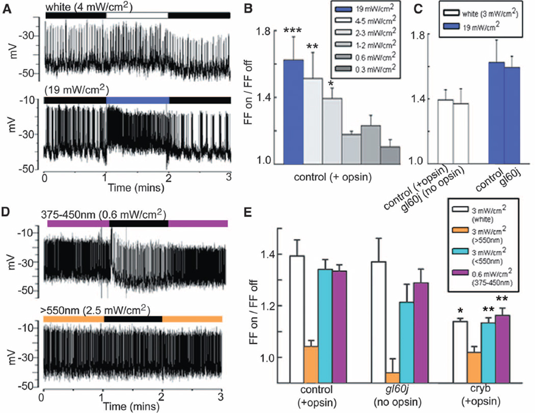
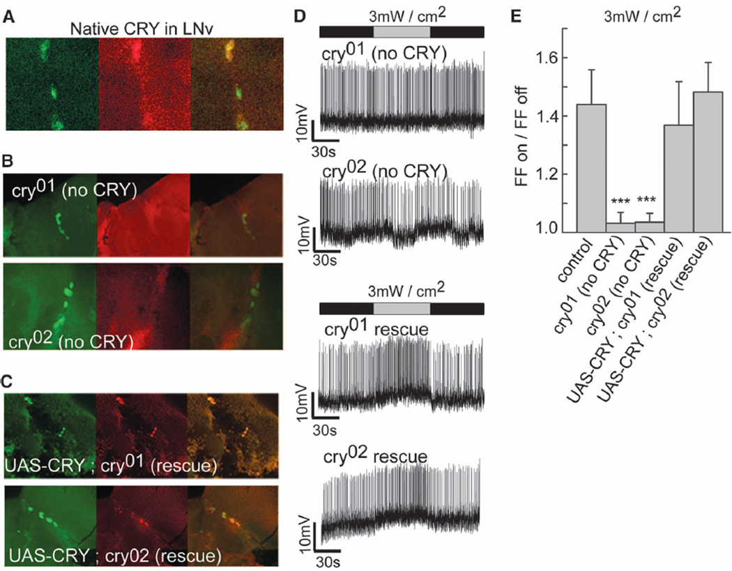
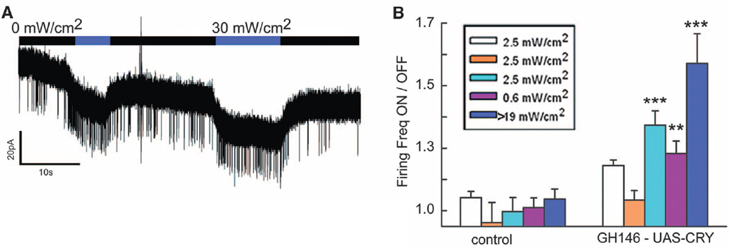
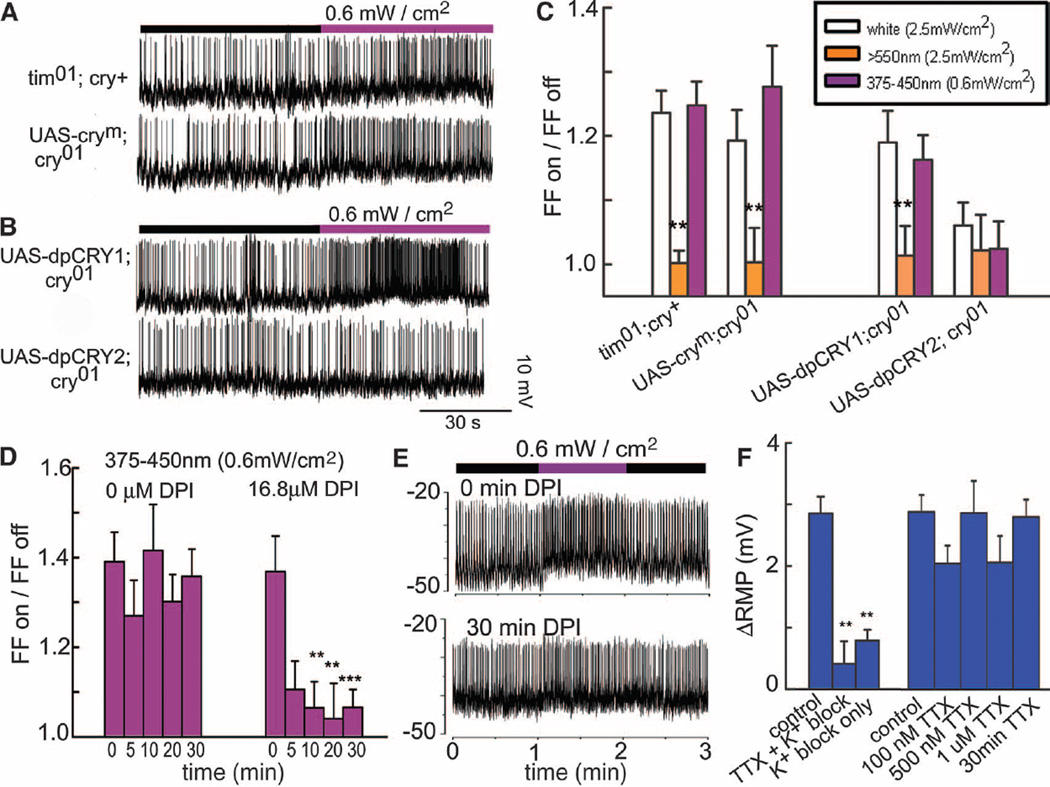
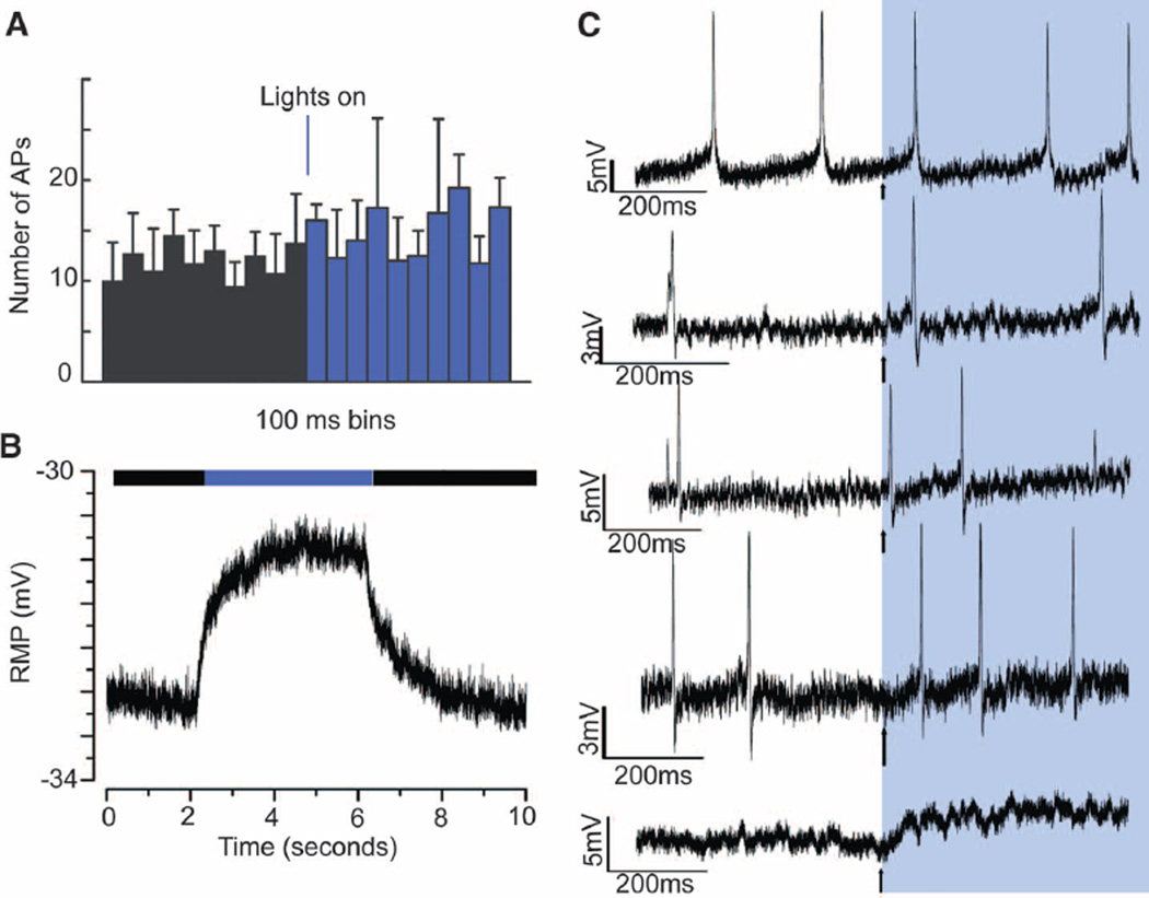
Light-responsive neural activity in central brain neurons is generally conveyed through opsin-based signaling from external photoreceptors. Large lateral ventral arousal neurons (lLNvs) in Drosophila melanogaster increase action potential firing within seconds in response to light in the absence of all opsin-based photoreceptors. Light-evoked changes in membrane resting potential occur in about 100 milliseconds. The light response is selective for blue wavelengths corresponding to the spectral sensitivity of CRYPTOCHROME (CRY). cry-null lines are light-unresponsive, but restored CRY expression in the lLNv rescues responsiveness. Furthermore, expression of CRY in neurons that are normally unresponsive to light confers responsiveness. The CRY-mediated light response requires a flavin redox-based mechanism and depends on potassium channel conductance, but is independent of the classical circadian CRY-TIMELESS interaction.


Figures

Fig. 1
Fig. 1
Drosophila lLNvs rapidly increase spontaneous action potential firing rate in response to light independent of opsin-based classical photoreceptors. (A) (Top) Response of a representative tonic firing cell to 4 mW/cm2 halogen white light. (Bottom) Light response of burst firing cell to 19 mW/cm2 blue light. Alternating light/dark cycles denoted by white or blue versus black bars above traces. (B) Firing frequency in light/dark varies according to light intensity. *P ≤ 0.05, **P ≤ 0.01, ***P ≤ 0.005; error bars indicate SEM. (C) Genetic ablation of all external opsin-based photoreceptors has no effect on the lLNv light response. (D) Representative recordings of lLNv light response evoked by blue-violet and orange-red light. Alternating light/dark cycles denoted by violet, orange, and black bars. (E) Spectral profiles of light responses of lLNv from control versus eyeless-null gl60j mutant flies are indistinguishable, but responses to white and blue light of cryb hypomorphs are significantly reduced.
Fig. 2
Fig. 2
The lLNv light response is absent in cry-null flies but is functionally rescued by targeted expression of CRY in the LNvs. (A) Native CRY is detected by anti-CRY and colocalizes in the LNv to a cryGAL4-driven GFP signal. (B) Anti-CRY signal (red, middle) is absent in LNv (green pdfGAL4/UAS-dORK-NC1-GFP, left) in cry01 and cry02 null flies (overlay, right). (C) Verification of anti-CRY signal (red) expressed specifically in LNv (labeled in green, left, pdfGAL4/UAS-dORK-NC1-GFP) in cry01 and cry02 null flies that express CRY driven by pdfGAL4 (overlay, right). (D) Representative recordings of cry-null flies (top) and genetic rescue of the lLNv light response in cry-null flies (bottom). (E) lLNv FF response evoked by 3 mW/cm2 white light/dark does not differ between control and LNv specific expression of CRY in cry01 and cry02 null genetic background flies.
Fig. 3
Fig. 3
Ectopic expression of CRY in inherently light-insensitive neurons renders them light-responsive. (A) Representative voltage clamp recording of CRY-expressing olfactory projection neuron shows light response evoked by 30 mW/cm2 blue light. (B) Spectral profile of light responses of olfactory projection neurons recorded from control (left) versus CRY-expressing (right) neurons. Control olfactory projection neurons are nonresponsive to all light wavelengths and intensities tested, whereas CRY-expressing olfactory neurons increase their firing rate in response to white, blue-green, and blue-violet light but not to orange light.
Fig. 4
Fig. 4
The CRY-mediated electrophysiological light response membrane depolarization by potassium channel modulation depends on flavin-specific redox reactions rather than TIM interaction. (A) Representative recordings of lLNv expressing CRY but tim null (tim01, top) and cry01 with pdfGAL4-driven CRYM (CRYM, bottom). (B) Recordings from lLNvs expressing UAS-driven D. plexippus dpCRY1 (top) or dpCRY2 exposed to blue-violet (purple bar) after darkness (black bar). (C) White and violet light evoke significant responses from each genotype except dpCRY2; cry01. (D) Treatment with the redox inhibitor DPI rapidly attenuates the light response. (E) Representative recordings of light-evoked responses in vehicle (top) versus 30 min of DPI treatment (bottom) for the same cell. (F) Light-evoked depolarization in lLNv is significantly decreased after treatment with potassium channel blockers TEA, 4-AP, and CsCl in the presence or absence of TTX.
Fig. 5
Fig. 5
RMP changes rapidly upon lights on and off. (A) Number of spikes in 100-ms bins shown for 1 s before and after the onset of an intense blue light pulse (~35 mW/cm2), averaged for 60 sweeps from four lLNvs. (B) The RMP shows an evoked increase upon triggering of the intense blue light. Trace is an average of 120 sweeps recorded from four lLNvs. The evoked rise and fall were fitted with double exponential functions. (C) Individual records that contributed to the averages depicted in (A) and (B). Four of five traces (bottom four traces) show an appreciable rise in RMP within 100 ms, but the increase in FF for the four cells that are firing is apparent only after several hundred milliseconds.

Comment in

  • Neuroscience. A CRY to rise.
    Im SH, Taghert PH. Im SH, et al. Science. 2011 Mar 18;331(6023):1394-5. doi: 10.1126/science.1204293. Science. 2011. PMID: 21415342 No abstract available.

Similar articles

Cited by

References

    1. Sheeba V, et al. Curr. Biol. 2008;18:1537. - PMC - PubMed
    1. Parisky KM, et al. Neuron. 2008;60:672. - PMC - PubMed
    1. Shang Y, Griffith LC, Rosbash M. Proc. Natl. Acad. Sci. U.S.A. 2008;105:19587. - PMC - PubMed
    1. Sheeba V, Gu H, Sharma VK, O’Dowd DK, Holmes TC. J. Neurophysiol. 2008;99:976. - PMC - PubMed
    1. Helfrich-Förster C, Stengl M, Homberg U. Chronobiol. Int. 1998;15:567. - PubMed

Publication types

MeSH terms