

Iron overload: accuracy of in-phase and out-of-phase MRI as a quick method to evaluate liver iron load in haematological malignancies and chronic liver disease
- PMID: 21385919
- PMCID: PMC3474105
- DOI: 10.1259/bjr/22327146
Iron overload: accuracy of in-phase and out-of-phase MRI as a quick method to evaluate liver iron load in haematological malignancies and chronic liver disease
Abstract
Objectives: The purpose of this prospective study was to evaluate the accuracy of in-phase and out-of-phase imaging to assess hepatic iron concentration in patients with haematological malignancies and chronic liver disease.
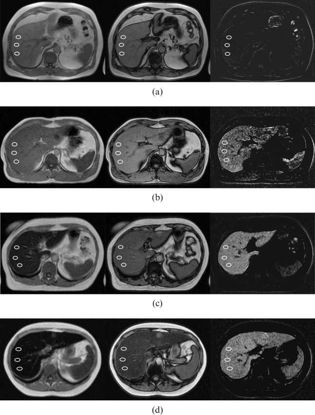
Methods: MRI-based hepatic iron concentration (M-HIC, μmol g(-1)) was used as a reference standard. 42 patients suspected of having iron overload and 12 control subjects underwent 1.5 T in- and out-of-phase and M-HIC liver imaging. Two methods, semi-quantitative visual grading made by two independent readers and quantitative relative signal intensity (rSI) grading from the signal intensity differences of in-phase and out-of-phase images, were used. Statistical analyses were performed using the Spearman and Kruskal-Wallis tests, receiver operator curves and κ coefficients.
Results: The correlations between M-HIC and visual gradings of Reader 1 (r = 0.9534, p < 0.0001) and Reader 2 (r = 0.9456, p < 0.0001) were higher than the correlations of the rSI method (r = 0.7719, p < 0.0001). There was excellent agreement between the readers (weighted κ = 0.9619). Both visual grading and rSI were similar in detecting liver iron overload: rSI had 84.85% sensitivity and 100% specificity; visual grading had 85% sensitivity and 100% specificity. The differences between the grades of visual grading were significant (p < 0.0001) and the method was able to distinguish different degrees of iron overload at the threshold of 151 μmol g(-1) with 100% positive predictive value and negative predictive value.
Conclusion: Detection and grading of liver iron can be performed reliably with in-phase and out-of-phase imaging. Liver fat is a potential pitfall, which limits the use of rSI.
Figures
References
-
- Brittenham G, Badman D. Noninvasive measurement of iron: report of an NIDDK workshop. Blood 2003;101:15–9 - PubMed
-
- Andrews N. Disorders of iron metabolism. N Engl J Med 1999;341:1986–95 - PubMed
-
- Adams P. Review article: the modern diagnosis and management of haemochromatosis. Aliment Pharmacol Ther 2006;23:1681–91 - PubMed
-
- Alústiza J, Castiella A, De Juan M, Emparanza J, Artetxe J, Uranga M. Iron overload in the liver diagnostic and quantification. Eur J Radiol 2007;61:499–506 - PubMed
-
- Angelucci E, Brittenham G, McLaren C, Ripalti M, Baronciani D, Giardini C, et al. Hepatic iron concentration and total body iron stores in thalassemia major. N Engl J Med 2000;343:327–31 - PubMed
MeSH terms
Substances
LinkOut - more resources
Full Text Sources
Medical
