. 2011 May;152(5):2001-10.
doi: 10.1210/en.2010-0964. Epub 2011 Mar 8.

Androgenic influence on serotonergic activation of the HPA stress axis

Affiliations

Androgenic influence on serotonergic activation of the HPA stress axis

Nirupa Goel et al. Endocrinology. 2011 May.

Abstract

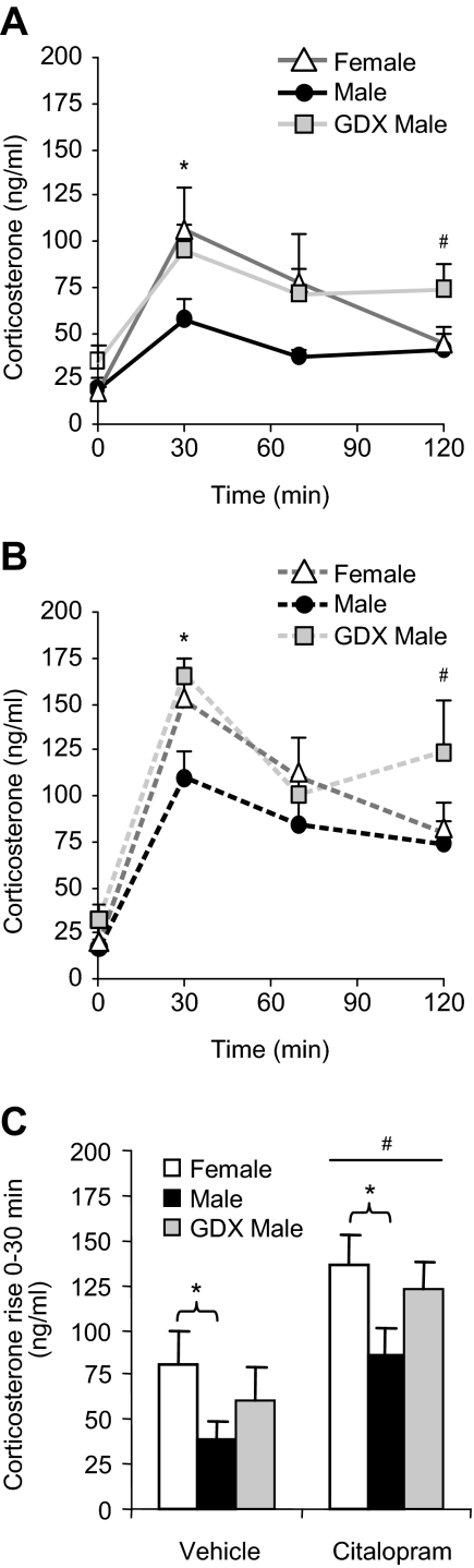
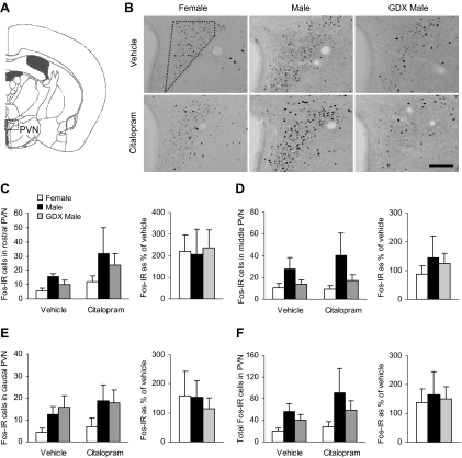
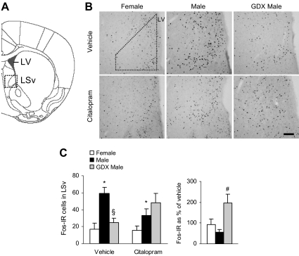
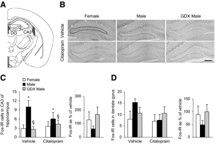
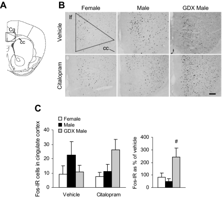
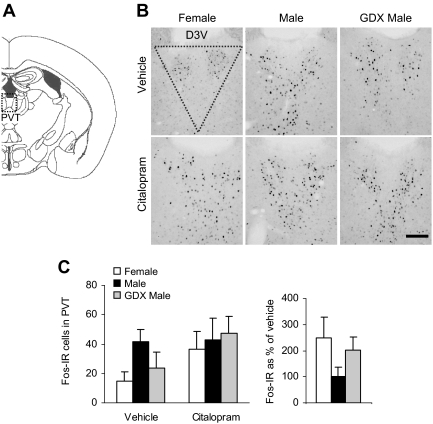
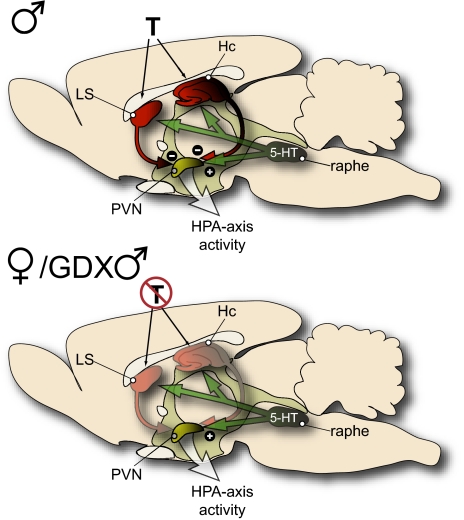
The higher incidence of stress-mediated affective disorders in women may be a function of gonadal hormone influence on complex interactions between serotonin and neural circuits that mediate the hypothalamic-pituitary-adrenal (HPA) stress axis. The paraventricular nucleus of the hypothalamus (PVN) receives serotonergic innervation, and selective serotonin reuptake inhibitors such as citalopram activate the HPA axis independent of stress. We have previously demonstrated that the magnitude of this serotonergic activation was greater in females and was attenuated by testosterone administration; however, the potential central sites of action where androgens reduce these serotonergic effects have not been determined. Therefore, we examined a time course of corticosterone production and used central c-Fos protein levels to assay neuronal activation in stress-related brain regions in female, male, and gonadectomized male mice after an acute citalopram injection (15 mg/kg). In the hippocampus, c-Fos-immunoreactivity was greater in males than in females or gonadectomized males. This same pattern emerged in the lateral septum after vehicle and gonadectomy reversed the effect of citalopram. These regions are important for inhibitory influences on the PVN, and accordingly, hippocampal c-Fos levels were negatively correlated with corticosterone production. No sex differences in c-Fos were detected in the PVN, cingulate cortex, or paraventricular thalamus in response to vehicle or citalopram. These data support brain region-specific regulation of the HPA axis where sex differences may be mediated partly through androgen enhancement of signaling in inhibitory regions.


Figures

Fig. 1.
Fig. 1.
Corticosterone levels in response to citalopram. Female, male, and GDX males were injected with vehicle (A) or citalopram (B), and plasma corticosterone was measured. Corticosterone levels were lower in males than in females and GDX males in both conditions. The difference was most notable at 30 min (*, P < 0.05), and we observed a delay in the recovery in GDX males (#, P < 0.05). C, Shows the corticosterone rise from 0–30 min, which was greater in females than in males after vehicle or citalopram administration (*, P < 0.05). We also found a main effect of citalopram on the initial rise of corticosterone (#, P < 0.001).
Fig. 2.
Fig. 2.
No differences between male and female mice were detected for c-Fos-IR in the PVN after vehicle or citalopram injection. A, Diagram of brain section used for PVN analysis, adapted from the mouse atlas (24). B, Representative images of c-Fos-IR in the middle PVN. Dotted shape indicates area analyzed. 3V, Third ventricle. Scale bar, 100 μm. C–E, c-Fos-IR in the rostral, middle, and caudal subregions of the PVN and the effect of citalopram expressed as a percentage of vehicle. F, Total c-Fos-IR across the rostrocaudal axis of the PVN and the response to citalopram expressed as a percentage of vehicle.
Fig. 3.
Fig. 3.
GDX reversed the effect of citalopram on c-Fos-IR in the LSv. A, Diagram of brain section used for LSv analysis, adapted from the mouse atlas (24). B, Representative images of c-Fos-IR in the LSv. Dotted shape indicates area analyzed. LV, Lateral ventricle. Scale bar, 100 μm. C, Male mice had more c-Fos-IR cells than females after vehicle or citalopram injections (*, P < 0.05). After vehicle treatment, c-Fos levels were lower in GDX males compared with intact males (§, P < 0.05), but this pattern was reversed after citalopram treatment. Citalopram increased c-Fos-IR only in GDX males (#, P < 0.05).
Fig. 4.
Fig. 4.
Intact males exhibited greater c-Fos-IR in the hippocampus than females or GDX males in response to vehicle or citalopram. A, Diagram of brain section used for dentate gyrus (DG) and CA3 hippocampal analysis, adapted from the mouse atlas (24). B, Representative images of c-Fos-IR in the DG (CA3 region not shown). Dotted shape indicates area analyzed. Scale bar, 200 μm. C, In the CA3 subfield of the hippocampus, intact males showed greater c-Fos-IR than females (*, P < 0.05) and GDX males (§, P < 0.01). D, c-Fos levels were similar between groups in the dentate gyrus. Citalopram did not significantly alter c-Fos levels compared with vehicle injection in the CA3 or dentate gyrus subregions.
Fig. 5.
Fig. 5.
c-Fos-IR in the cingulate cortex after vehicle or citalopram injection is similar in males, females, and GDX males. A, Diagram of brain section used for cingulate cortex (Cg) analysis, adapted from the mouse atlas (24). B, Representative images of c-Fos-IR in the Cg. Dotted shape indicates area analyzed. Scale bar, 100 μm. C, There were no group differences in c-Fos levels in the cingulate cortex. Citalopram increased c-Fos-IR only in GDX males (#, P < 0.05). cc, Corpus callosum; lf, longitudinal fissure.
Fig. 6.
Fig. 6.
No differences between males and females were detected in the c-Fos-IR response to vehicle or citalopram in the PVT. A, Diagram of brain section used for PVT analysis, adapted from the mouse atlas (24). B, Representative images of c-Fos-IR in the PVT. Dotted shape indicates area analyzed. Scale bar, 100 μm. D3V, Dorsal third ventricle. C, c-Fos levels in the PVT were similar across all groups and were not altered by citalopram injection.
Fig. 7.
Fig. 7.
Schematic image illustrating a hypothesized model explaining androgen-mediated differences in activation of the HPA stress axis. Equivalent levels of c-Fos-IR in the PVN of male, female, and GDX males suggest that neural activity in the PVN did not account for the observed differences in corticosterone levels after vehicle or 5-HT stimulation. Instead, the present data suggest that regions known to provide negative feedback to the HPA axis, such as the LS and hippocampus (Hc), are more active in the presence of androgen (T) but not in its absence. Accordingly, we hypothesize that androgens modulate the stress circuitry by enhancing inhibitory signaling to the PVN.

Similar articles

Cited by

References

    1. Kendler KS, Karkowski LM, Prescott CA. 1999. Causal relationship between stressful life events and the onset of major depression. Am J Psychiatry 156:837–841 - PubMed
    1. Kessler RC, Berglund P, Demler O, Jin R, Merikangas KR, Walters EE. 2005. Lifetime prevalence and age-of-onset distributions of DSM-IV disorders in the National Comorbidity Survey Replication. Arch Gen Psychiatry 62:593–602 - PubMed
    1. Arborelius L, Owens MJ, Plotsky PM, Nemeroff CB. 1999. The role of corticotropin-releasing factor in depression and anxiety disorders. J Endocrinol 160:1–12 - PubMed
    1. Bale TL. 2005. Sensitivity to stress: dysregulation of CRF pathways and disease development. Horm Behav 48:1–10 - PubMed
    1. Holden C. 2005. Sex and the suffering brain. Science 308:1574. - PubMed

Publication types

MeSH terms