Review
. 2011 Dec;54(6):1709-31.
doi: 10.1044/1092-4388(2011/10-0149). Epub 2011 Mar 8.

Biotechnology in the treatment of sensorineural hearing loss: foundations and future of hair cell regeneration

Affiliations
Review

Biotechnology in the treatment of sensorineural hearing loss: foundations and future of hair cell regeneration

Mark A Parker. J Speech Lang Hear Res. 2011 Dec.

Abstract

Purpose: To provide an overview of the methodologies involved in the field of hair cell regeneration. First, the author provides a tutorial on the biotechnological foundations of this field to assist the reader in the comprehension and interpretation of the research involved in hair cell regeneration. Next, the author presents a review of stem cell and gene therapy and provides a critical appraisal of their application to hair cell regeneration. The methodologies used in these approaches are highlighted.

Method: The author conducted a narrative review of the fields of cellular, molecular, and developmental biology, tissue engineering, and stem cell and gene therapy using the PubMed database.

Results: The use of biotechnological approaches to the treatment of hearing loss--approaches such as stem cell and gene therapy-has led to new methods of regenerating cochlear hair cells in mammals.

Conclusions: Incredible strides have been made in assembling important pieces of the puzzle that comprise hair cell regeneration. However, mammalian hair cell regeneration using stem cell and gene therapy are years--if not decades--away from being clinically feasible. If the goals of the biological approaches are met, these therapies may represent future treatments for hearing loss.


Figures

Figure 1
Figure 1. Gross Anatomy of the Mammalian Cochlea
A) The cochlea is composed of many cell types including bone (purple), ligament, sensory epithelia, and nerve that enables communication between the sensory epithelia and the brain. B) Close-up of the sensory epithelium (boxed region in A) illustrates the organ of Corti, which consists of inner and outer hair cells and their corresponding supporting cells. Hair cells are responsible for transducing the mechanical motion of the basilar membrane into neural impulses that can be interpreted as sound by the brain. The supporting cells act to provide structural and nutritional support for hair cells and possibly act as hair cell progenitors as well. C) Ototoxic damage, such as exposure to noise or aminoglycoside antibiotics, results in hair cell loss and subsequent hearing loss. In some instances, the supporting cells remain intact after damage and may act as a target for gene therapy. D) It is also possible for ototoxic damage to completely ablate the organ of Corti. In such an instance, the remaining cells form a flat epithelium that spans the basilar membrane.
Figure 3
Figure 3. Cellular and Molecular Biology in Hearing Research
Gene expression requires a molecular signal to initiate the transcription of DNA into an RNA molecule and then translation of the RNA into a protein. Common assays used to measure these cellular processes include the generation of transgenic animals, in situ hybridization, RT-PCR, and Western blot analysis. See text for details.
Figure 2
Figure 2. Molecular Genetics in Hearing Research
A) Gene is expression is tightly regulated. Typically, genes are quiescent and not transcribed until transcription factors bind to a region of DNA call a promoter (B). Each gene has a specific promoter and several factors may be required to bind to the promoter before transcription of the gene into RNA may begin (yellow arrow). C) A common method to measure gene expression is to insert an easily detectable gene, such as the gene for GFP, into the DNA just after the gene-of-interest. In this model, the GFP gene will be transcribed and then translated into protein at the same time as the gene-of-interest. The GFP protein is easily detected using a fluorescent microscope. D) In some cases, such as when the control of gene expression is to be measured, the molecular tag is inserted into the DNA after the promoter rather than after the gene-of-interest. E) In a gene knock-out organism, the gene-of-interest has been deleted or otherwise mutated to render it dysfunctional. Sometimes, the gene-of-interest is deleted and replaced by a molecular marker such as GFP (D).
Figure 4
Figure 4. Development of the murine cochlea
A) Cochlear development starts at approximately E8.5 with a thickening of a group of cells on the lateral wall of the developing fetus called the otic placode (blue). The cells of the otic placode invaginate (B) and pinch off from the surface of the embryo to form the otocyst by E10 (C). The red line in A indicates the cross section illustrated in B and C. D) Experiments where latex paint was injected into the developing scala media illustrate the development of the inner ear. The dorsal portion of the otocyst develops into the vestibular system (v) and the ventral portion develops into the cochlea (c). The scala media of the cochlea begins to elongate from the vestibule and spirals apically. Images adapted with permission from Morsli et al., (1998); Society for Neuroscience Publisher. E) Cross section through the E12 otocyst (left dashed line in D) highlights a group of cells that immunolabel for the Jagged-1 protein (Jag1; green), which labels the prosensory domain in the developing cochlea which will develop into the organ of Corti. F) Cross section through the postnatal day 1 (P1) organ of Corti (right dashed line in D) shows most of the adult cells are present at birth, including hair cells that immunolabel for the myosin 7a protein (green). However, the tunnel of Corti has not developed and Kölliker's organ (k) remains in place of the inner sulcus. G) The prosensory domain, identified by jag1 and sox2 expression, begins to express p27kip1 between E12 and E16 which initiates maturation of the sensory epithelium (H). I) Hair cell development begins within the prosensory domain at E13 when hair cells express atoh1 and signal their surrounding cells to differentiate into supporting cells. J) By E18, most of the supporting cells types are present in the sensory epithelium, however; the auditory system is not fully developed into 2 weeks after birth in mice.
Figure 5
Figure 5. Spontaneous Regeneration of Auditory Hair Cells the Chick
LEFT PANEL. A) Unlike the mammalian organ of Corti, the auditory sensory epithelium in the chick is covered in many rows of hair cells. B) Exposure to loud sound results in significant damage to sensory epithelia including loss of hair cells. C, E and F) Small ciliated cells spontaneously appear on the surface of the epithelium by six days after noise exposure. These cells maintain morphological similarities to developing hair cells. D) A significant number of hair cells appear on the epithelia by 10 days after the noise damage. These cells appear morphologically similar to adult hair cells with the exception that the orientations of their cilia are often askew. Image taken from Corwin and Cotanche (1988) reprinted with permission from AAAS and authors. RIGHT PANEL. The current model of hair cell regeneration holds that there are two mechanisms of hair cell regeneration in chicks (Stone and Cotanche, 2007). In each of these models, hair cells are regenerated from supporting cells that survived the ototoxic stimulus (A, B). C) Initially, supporting cells differentiate into a hair cell in a process called direct transdifferentiation. D) Later in the regenerative process, supporting cells enter into the mitotic cycle and undergo cell division. Of the two daughter cells, one will regenerate into a supporting cell and one will differentiate into a regenerated hair cell.
Figure 6
Figure 6. Plasticity in the Mammalian Organ of Corti
The ability of the mammalian cochlea to produce hair cells after normal cochlear development suggests that the organ of Corti maintains the proper cell types required for hair cell regeneration. A) In the mouse cochlea, hair cells are normally developed by 14 days after fertilization. Red cell=one row of inner hair cells, blue cells = three rows of outer hair cells. B) Culturing the organs of Corti in the presence of retinoic acid, a mitotic agent that induces cellular proliferation, results in the production of supernumerary hair cells (Kelley et al., 1993).
Figure 7
Figure 7. Stem Cell Replacement Therapy in the Cochlea
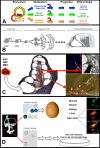
A) Pluripotent ESCs are derived from the developing blastocyst and have the capacity to differentiate into any cell type. When these cells divide, the daughter cells maintain the capacity for self renewal (curved arrow) and the potential to develop into any cell type within the developing fetus. ASCs, such as mesenchymal, neural, and intestinal stem cells, are multipotent cells found in the end-organs of adult tissue that maintain the ability for self renewal and produce daughter cells that can differentiate into many of the cell types that comprise the organ in which they reside. By contrast, progenitor cells exhibit a more limited potential and differentiate into a restricted number of cell types. B) The goals of stem cell replacement therapy are to use ESC, ASCs, or cochlear progenitor cells to replace dead or damaged cell types which result in hearing loss. C) Initial experimenters aiming to achieve this goal injected various types of stem cells into the deafened cochlea and later examined the differentiated fates of the transplanted stem cells. As discussed in the text, most stem cells types were able to survive transplantation and engraft into the cochlea, but the terminal cell type and their degree of differentiation depended on the specific type of injected stem cell. Images adapted with permission from TOP LEFT ((Shi et al., 2007) John Wiley and Sons; TOP RIGHT (Corrales et al., 2006) John Wiley and Sons; BOTTOM (Parker et al., 2007) Elsevier. D) Investigators are trying to determine whether the mammalian inner ear may harbor a population of dormant stem cells. Li et al (2003) isolated cells with stem cell properties from the developing vestibular system (vestibular spheres), injected them into a chicken egg, incubated the egg, and then assayed for terminal differentiation of the transplanted cells (which expressed GFP). The authors suggest that the vestibular spheres are pluripotent stem cells because they differentiated into many different tissue types, including hair cells. Reprinted by permission from Macmillan Publishers Ltd: Nature Medicine, (Li et. al. 2003), copyright (2003). In addition, several laboratories are investigating the ability of similar cochlear spheres to differentiate into hair cells in vitro.
Figure 8
Figure 8. Gene Delivery in Hearing Research
TOP PANEL. Viral mediated gene delivery utilizes virus particles to deliver a gene-of-interest into a cell. A) A virus (blue) attaches to a cell (B). Once attached, the DNA of the virus (black) will be inserted into the cytoplasm of the host cell (C-D) where the viral DNA will incorporate into the DNA of the host cell (red) (E). Viral DNA consists of self-replicating genes and will cause the host cell to manufacture more viral particles (F). G) Eventually, the host cell will release vial particles back into the environment. H) In a viral-mediated gene delivery system, the viral DNA that codes for self-replication is replaced with a gene-of-interest, such as atoh1. Cells that are infected with the engineered virus will express the atoh1. Image adapted from (Stewart, Fuller, & Burnett, 1993). MIDDLE PANEL. Izumikawa et al. (2005) injected an adenovirus that carried the atoh1 gene into the cochleas of chemically deafened adult guinea pigs. The results indicated that the guinea pigs injected with the atoh1 gene had better ABR threshold and more hair cells than controls injected with virus particles lacking the atoh1 gene. Arrow heads in the far right panel highlight extra streocilia bundles. Asterisk marks the site of injection into the scala media. Adapted by permission from Macmillan Publishers Ltd: Nature Neuroscience (Izumikawa et al., 2005), copyright (2005). BOTTOM PANEL. Genes can also be delivered into the inner ear using pulse trains of electric charge (electroporation). Gubbels et al. (2008) injected atoh1 DNA into the developing otocysts of E11.5 mice in utero, electroporated the embryos, allowed them to mature, and then analyzed the cochleas at various time points for hair cell markers. These results indicated that the overexpression of atoh1 during embryogenesis resulted in extra rows of functional hair cells into adulthood. Arrows in far right point to hair cells that exhibit abnormal stereocilia. Reprinted by permission from Macmillan Publishers Ltd: Nature (Gubbels et al., 2008), copyright (2008).
Figure 9
Figure 9. Expression of the atoh1 Gene in the organ of Corti is Sufficient for Hair Cell Genesis
A) ISH shows that hair cells express RNA for atoh1 gene (black) by E17. Arrow indicates Deiters’ cell layer. Image adapted from (Lanford, Shailam, Norton, Ridley, & Kelley, 2000). B and C) Organs of Corti that were isolated from perinatal rats, then electroporated with the atoh1 gene tagged with a GFP marker (green) expressed the hair cell marker myosin 7a (red) when assayed by immunohistochemestry, indicating that these cells expressed hair cell specific markers. Most of the regenerated hair cells were seen in Kölliker's organ (a.k.a. the greater epithelial ridge, (GER)). Image in C is the overlay of B and both were adapted by permission from Macmillan Publishers Ltd: Nature Neuroscience (Zheng & Gao, 2000), copyright (2000). D) Adult guinea pigs were deafened, and then their cochleas were infected with a virus that had been engineered to deliver atoh1. Numerous regenerated hair cells appeared in the infected cochleas. In some cases, the regenerated hair cells maintained the morphology of ciliated supporting cells (nucleus close to basilar membrane with a cellular process that reached the reticular lamina). White arrow: basilar membrane; black arrow: cilia, black arrowhead: reticular lamina. Adapted by permission from Macmillan Publishers Ltd: Nature Neuroscience (Izumikawa et al., 2005), copyright (2005). E) Viral-mediated delivery of atoh1 results in random infection of cells within the cochlea. Each colored cell nucleus indicates a cell type that exhibited a hair cell phenotype after infection of atoh1 into the deafened cochlea. Image adapted with permission from Kawamoto et al. (2003); Society for Neuroscience Publisher.

Similar articles

Cited by

References

    1. Abello G, Alsina B. Establishment of a proneural field in the inner ear. Int J Dev Biol. 2007;51(6-7):483–493. - PubMed
    1. Akazawa C, Ishibashi M, Shimizu C, Nakanishi S, Kageyama R. A mammalian helix-loop-helix factor structurally related to the product of Drosophila proneural gene atonal is a positive transcriptional regulator expressed in the developing nervous system. J Biol Chem. 1995;270(15):8730–8738. - PubMed
    1. Alberts B, Johnson A, Lewis J, Raff M. Molecular Biology of the Cell. 5 ed. Garland Science; New York: 2007.
    1. Alford BR, Ruben RJ. Physiological, behavioral and anatomical correlates of the development of hearing in the mouse. Ann Otol Rhinol Laryngol. 1963;72:237–247. - PubMed
    1. Alibardi L. Morphological and cellular aspects of tail and limb regeneration in lizards. A model system with implications for tissue regeneration in mammals. Adv Anat Embryol Cell Biol. 2010;207:iii, v–x, 1–109. - PubMed

Publication types

MeSH terms