. 2011 Feb 28;6(2):e16722.
doi: 10.1371/journal.pone.0016722.

Cigarette smoke-related hydroquinone dysregulates MCP-1, VEGF and PEDF expression in retinal pigment epithelium in vitro and in vivo

Affiliations

Cigarette smoke-related hydroquinone dysregulates MCP-1, VEGF and PEDF expression in retinal pigment epithelium in vitro and in vivo

Marianne Pons et al. PLoS One. .

Abstract

Background: Age-related macular degeneration (AMD) is the leading cause of legal blindness in the elderly population. Debris (termed drusen) below the retinal pigment epithelium (RPE) have been recognized as a risk factor for dry AMD and its progression to wet AMD, which is characterized by choroidal neovascularization (CNV). The underlying mechanism of how drusen might elicit CNV remains undefined. Cigarette smoking, oxidative damage to the RPE and inflammation are postulated to be involved in the pathophysiology of the disease. To better understand the cellular mechanism(s) linking oxidative stress and inflammation to AMD, we examined the expression of pro-inflammatory monocyte chemoattractant protein-1 (MCP-1), pro-angiogenic vascular endothelial growth factor (VEGF) and anti-angiogenic pigment epithelial derived factor (PEDF) in RPE from smoker patients with AMD. We also evaluated the effects of hydroquinone (HQ), a major pro-oxidant in cigarette smoke on MCP-1, VEGF and PEDF expression in cultured ARPE-19 cells and RPE/choroids from C57BL/6 mice.

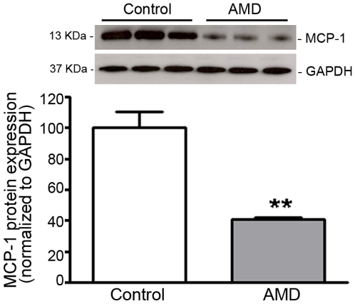
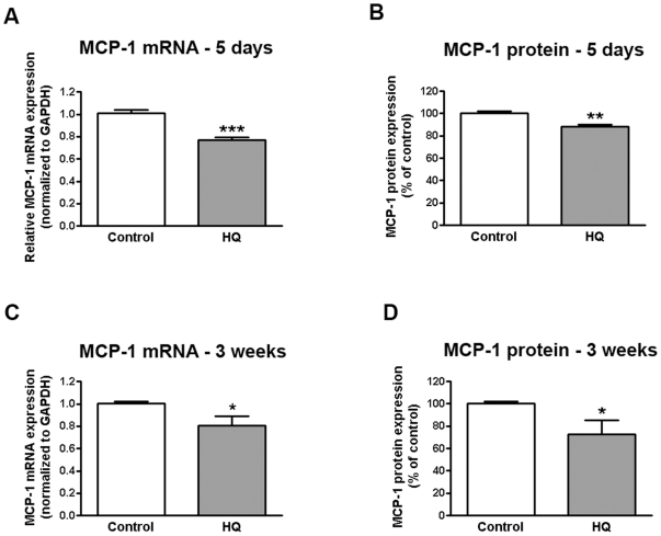
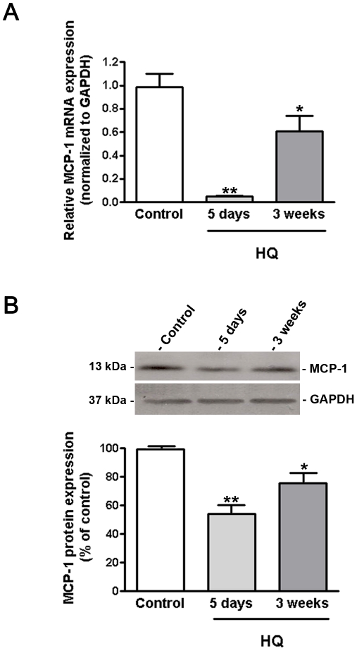
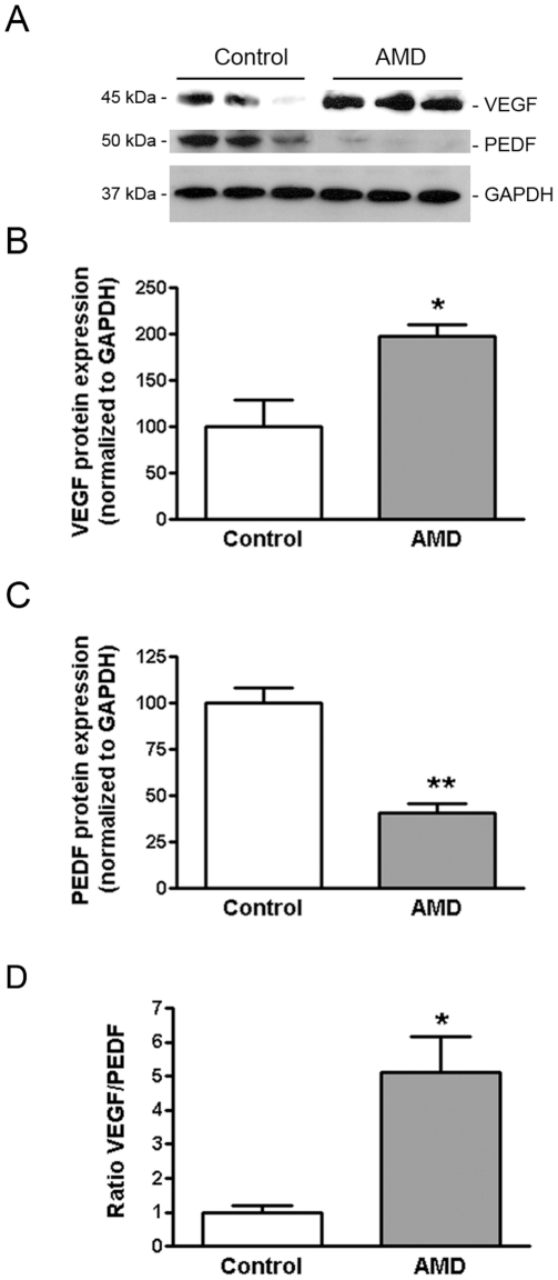
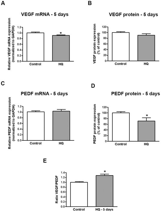
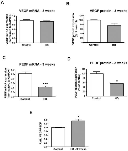
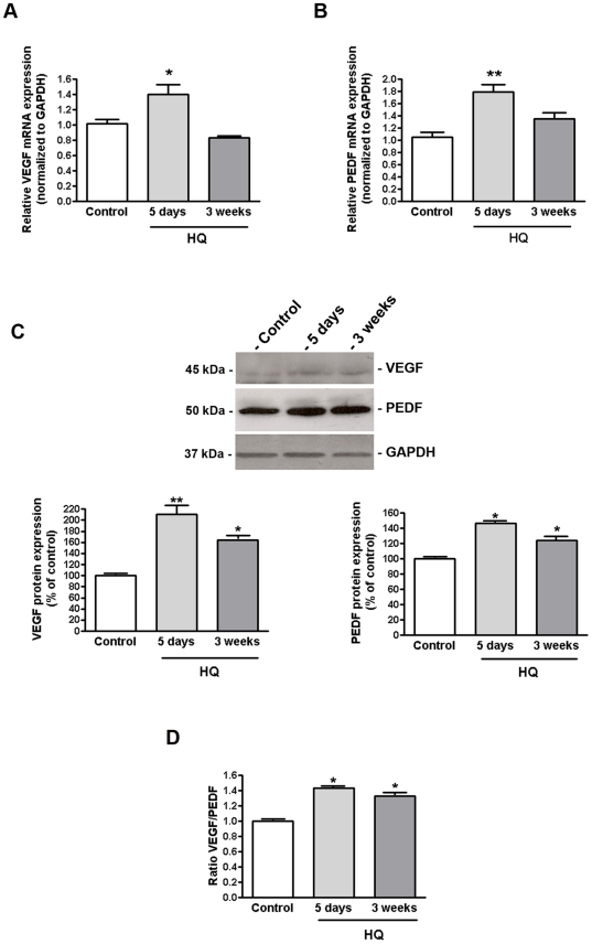
Principal findings: MCP-1, VEGF and PEDF expression was examined by real-time PCR, Western blot, and ELISA. Low levels of MCP-1 protein were detected in RPE from AMD smoker patients relative to controls. Both MCP-1 mRNA and protein were downregulated in ARPE-19 cells and RPE/choroids from C57BL/6 mice after 5 days and 3 weeks of exposure to HQ-induced oxidative injury. VEGF protein expression was increased and PEDF protein expression was decreased in RPE from smoker patients with AMD versus controls resulting in increased VEGF/PEDF ratio. Treatment with HQ for 5 days and 3 weeks increased the VEGF/PEDF ratio in vitro and in vivo.

Conclusion: We propose that impaired RPE-derived MCP-1-mediated scavenging macrophages recruitment and phagocytosis might lead to incomplete clearance of proinflammatory debris and infiltration of proangiogenic macrophages which along with increased VEGF/PEDF ratio favoring angiogenesis might promote drusen accumulation and progression to CNV in smoker patients with dry AMD.


Conflict of interest statement

Competing Interests: The authors have declared that no competing interests exist.

Figures

Figure 1
Figure 1. MCP-1 expression is decreased in RPE from smoker AMD patients.
MCP-1 protein expression was evaluated by Western blot in RPE lysates from 3 smoker donors with AMD and 3 age-matched non smoker controls with no known history of eye disease. GAPDH served as loading control. Top: representative Western blots of the indicated proteins. The numbers to the left are molecular weights in kilodaltons (KDa). Bottom: average densitometry results. Data are expressed as percentage of control and are means ± SE. **p<0.01 versus control.
Figure 2
Figure 2. Sustained and repetitive HQ-induced oxidative injury decreases on MCP-1 expression in human RPE cells.
Confluent serum-starved ARPE-19 cells were treated with (A, B) 10 µM HQ every 24 hours for 5 consecutive days or (C, D) 50 µM HQ every 4 days for 24 hours for 3 consecutive weeks in phenol red-free 0.1% FBS medium. Total RNA was extracted to assess MCP-1 mRNA expression by real-time PCR (A, C). GAPDH was used as internal control. Supernatants were collected to assess MCP-1 protein concentration by ELISA (B, D). Data are mean± SE and represent the average results of 3 independent experiments run in duplicate. * is p<0.05, ** is p<0.01 and *** is p<0.001 versus control.
Figure 3
Figure 3. MCP-1 expression is decreased in RPE/choroids from mice exposed to HQ.
(A) MCP-1 mRNA expression was downregulated in response to HQ-induced oxidative injury. Total RNA was extracted from microdissected RPE/choroid complexes after 5 days and 3 weeks of exposure to HQ in drinking water (0.8%). MCP-1 mRNA expression was measured by real-time PCR. GAPDH was used as internal control (n = 5 eyes per group). (B) MCP-1 protein expression was downregulated in response to HQ-induced oxidative injury. Total protein was extracted from microdissected RPE/choroid complexes after 5 days and 3 weeks of exposure to HQ in drinking water (0.8%). Equivalent amounts of protein from 5 eyes per group were pooled for each lane. MCP-1 protein expression was evaluated by Western blot and normalized to GAPDH. Top: representative Western blot gel. The numbers to the left are molecular weights in kilodaltons (KDa). Bottom: average densitometry results. Data are expressed as percentage of control and are means ± SE. * is p<0.05 and **p<0.01 versus control.
Figure 4
Figure 4. VEGF expression is increased and PEDF expression decreased in RPE from AMD patients.
VEGF and PEDF protein expression was evaluated by Western blot in RPE lysates from 3 smoker donors with AMD and 3 non smoker controls with no known history of eye disease. GAPDH served as loading control. (A) Representative Western blots of the indicated proteins. The numbers to the left are molecular weights in kilodaltons (KDa). (B, C) Average densitometry results. (D) VEGF-to-PEDF protein ratio. Data are expressed as percentage of control and are means ± SE. * is p<0.05 and **p<0.01 versus control.
Figure 5
Figure 5. Increased VEGF-to-PEDF ratio in human RPE cells following sustained oxidative injury with HQ.
Confluent serum-starved ARPE-19 cells were treated with 10 µM HQ every 24 hours for 5 consecutive days in phenol red-free 0.1% FBS medium. Total RNA was extracted to assess (A) VEGF and (D) PEDF mRNA expression by real-time PCR. GAPDH was used as internal control. Supernatants were collected to assess (B) VEGF and (D) PEDF protein concentration by ELISA. (E) VEGF-to-PEDF protein ratio. Data are mean± SE and represent the average results of 3 independent experiments run in duplicate. * is p<0.05 versus control.
Figure 6
Figure 6. Increased VEGF-to-PEDF ratio in human RPE cells following repetitive oxidative injury with HQ.
Confluent serum-starved ARPE-19 cells were treated with 50 µM HQ every 4 days for 24 hours for 3 consecutive weeks in phenol red-free 0.1% FBS medium. Total RNA was extracted to assess (A) VEGF and (D) PEDF mRNA expression by real-time PCR. GAPDH was used as internal control. Supernatants were collected to assess (B) VEGF and (D) PEDF protein concentration by ELISA. (E) VEGF-to-PEDF protein ratio. Data are mean± SE and represent the average results of 3 independent experiments run in duplicate. * is p<0.05 and *** is p<0.0001 versus control.
Figure 7
Figure 7. VEGF-PEDF balance is altered in RPE/choroids from mice exposed to HQ.
(A) VEGF and (B) PEDF mRNA expression in response to HQ-induced oxidative injury. Total RNA was extracted from microdissected RPE/choroid complexes after 5 days and 3 weeks of exposure to HQ in drinking water (0.8%). VEGF and PEDF mRNA expression was measured by real-time PCR. GAPDH was used as internal control (n = 5 eyes per group). (C) VEGF and PEDF protein expression in response to HQ-induced oxidative injury. (D) VEGF-to-PEDF protein ratio. Total protein was extracted from microdissected RPE/choroid complexes after 5 days and 3 weeks of exposure to HQ in drinking water (0.8%). Equivalent amounts of protein from 5 eyes per group were pooled for each lane. VEGF and PEDF protein expression was evaluated by Western blot and normalized to GAPDH. Top: representative Western blots of the indicated proteins. The numbers to the left are molecular weights in kilodaltons (KDa). Bottom: average densitometry results. Data are expressed as percentage of control and are means ± SE. * is p<0.05 and **p<0.01 versus control.

Similar articles

Cited by

References

    1. Augood CA, Vingerling JR, de Jong PT, Chakravarthy U, Seland J, et al. Prevalence of age-related maculopathy in older Europeans: the European Eye Study (EUREYE). Arch Ophthalmol. 2006;124:529–535. - PubMed
    1. Evans JR. Risk factors for age-related macular degeneration. Prog Retin Eye Res. 2001;20:227–253. - PubMed
    1. Javitt JC, Zhou Z, Maguire MG, Fine SL, Willke RJ. Incidence of exudative age-related macular degeneration among elderly Americans. Ophthalmology. 2003;110:1534–1539. - PubMed
    1. Klein R, Peto T, Bird A, Vannewkirk MR. The epidemiology of age-related macular degeneration. Am J Ophthalmol. 2004;137:486–495. - PubMed
    1. Friedman DS, O'Colmain BJ, Munoz B, Tomany SC, McCarty C, et al. Prevalence of age-related macular degeneration in the United States. Arch Ophthalmol. 2004;122:564–572. - PubMed

Publication types

MeSH terms