

Tangential vitreous traction: a possible mechanism of development of cystoid macular edema in retinitis pigmentosa
- PMID: 21386918
- PMCID: PMC3046995
- DOI: 10.2147/OPTH.S16891
Tangential vitreous traction: a possible mechanism of development of cystoid macular edema in retinitis pigmentosa
Abstract
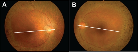
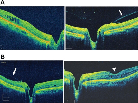
We report the possible mechanism of development of cystoid macular edema (CME) in retinitis pigmentosa (RP) in the case of a 68-year-old woman with RP and CME in the right eye and resolving CME in the left eye. Spectral domain optical coherence tomography showed CME and posterior vitreoschisis in the nasal quadrant of the fundus without a posterior vitreous detachment (PVD). This vitreous pathology suggested bilateral thickening and shrinkage of the posterior vitreous cortex. In the right eye, CME was evident with no vitreofoveal separation. However, in the left eye, minimal change was seen in the CME associated with a focal shallow PVD over the fovea. The best-corrected visual acuity (BCVA) in the left eye increased to 0.3 from 0.15 7 years after the first visit. Tangential vitreous traction on the macula may have caused the CME in the right eye. The shallow PVD over the fovea might have released the tangential vitreous traction from the fovea, induced spontaneous resolution of the CME, and improved the BCVA in the left eye.
Keywords: cystoid macular edema; optical coherence tomography; posterior vitreoschisis; posterior vitreous detachment; retinitis pigmentosa.
Figures
References
-
- Fetkenhour CL, Choromokos E, Weinstein J, Shoch D. Cystoid macular edema in retinitis pigmentosa. Trans Sect Ophthalmol Am Acad Ophthalmol Otolaryngol. 1977;83(3 Pt 1):OP515–OP521. - PubMed
-
- Newsome DA. Retinal fluorescein leakage in retinitis pigmentosa. Am J Ophthalmol. 1986;101(3):354–360. - PubMed
-
- Fishman GA, Fishman M, Maggiano J. Macular lesions associated with retinitis pigmentosa. Arch Ophthalmol. 1977;95(5):798–803. - PubMed
-
- Fishman GA, Maggiano JM, Fishman M. Foveal lesions seen in retinitis pigmentosa. Arch Ophthalmol. 1977;95(11):1993–1996. - PubMed
-
- Hirakawa H, Iijima H, Gohdo T, Tsukahara S. Optical coherence tomography of cystoid macular edema associated with retinitis pigmentosa. Am J Ophthalmol. 1999;128(2):185–191. - PubMed
Publication types
LinkOut - more resources
Full Text Sources
