

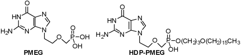
Antiproliferative property of hexadecyloxypropyl 9-[2-(phosphonomethoxy) ethyl] guanine (HDP-PMEG) for unwanted ocular proliferation
- PMID: 21386925
- PMCID: PMC3049735
Antiproliferative property of hexadecyloxypropyl 9-[2-(phosphonomethoxy) ethyl] guanine (HDP-PMEG) for unwanted ocular proliferation
Abstract
Purpose: To investigate the safety and inhibitory effects of hexadecyloxypropyl 9-[2-(phosphonomethoxy) ethyl] guanine (HDP-PMEG) on ocular cell proliferation and collagen matrix contraction.
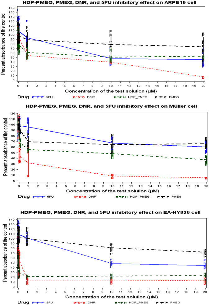
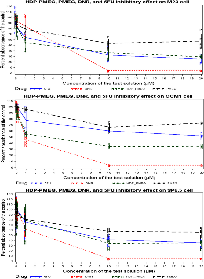
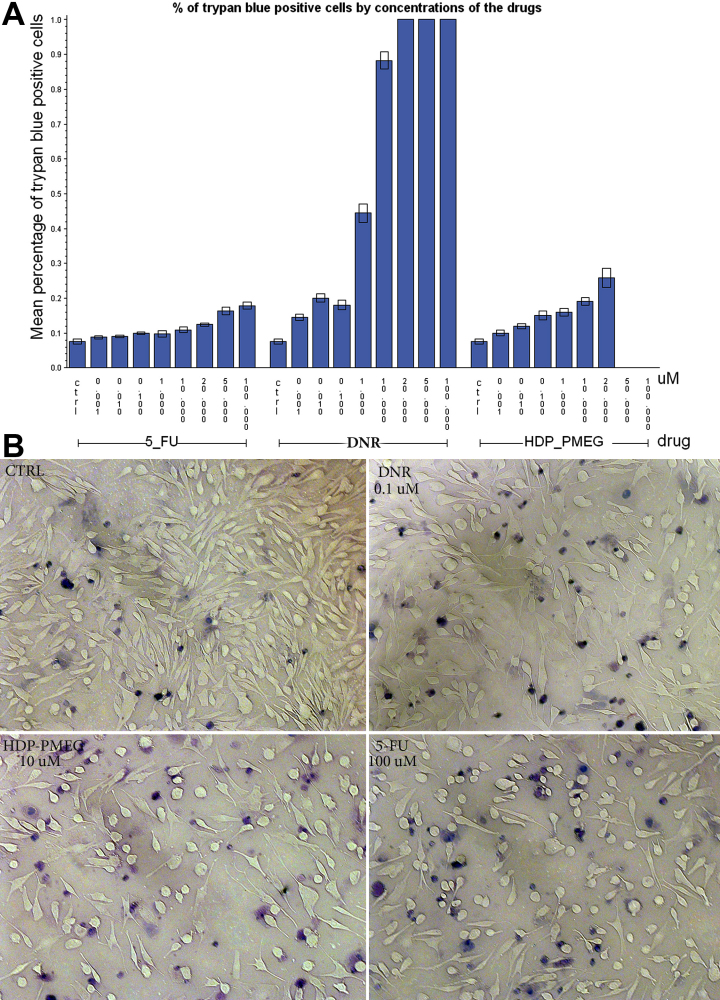
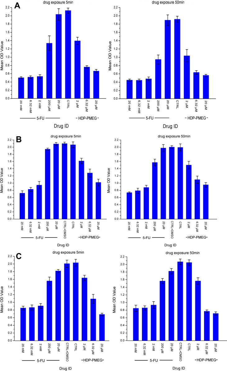
Methods: For the antiproliferation studies, various ocular cell monolayers were exposed to HDP-PMEG, PMEG, 5-fluorouracil (5-FU), and daunorubicin (DNB). For the collagen contraction studies, retinal pigment epithelium (RPE) cells seeded onto type I collagen lattices were exposed for a single 5- or 50-min period to various concentrations of HDP-PMEG or 5-FU. For the cytotoxicity study, trypan blue exclusion tests were performed using a human Müller cell line. Cytotoxicity was determined up to 4 days after treatment.
Results: The proliferation of RPE cells, scleral fibroblasts, vessel endothelial cells, and ocular melanoma cells can all be significantly inhibited by HDP-PMEG. Its inhibitory effects on those cells were uniformly stronger than that of 5-FU. Contraction of the collagen matrix containing RPE cells was significantly inhibited by HDP-PMEG and by 5-FU at concentrations of 20 µM and 2,000 µM, respectively, as compared with controls (p<0.05). The safety profile of HDP-PMEG was significantly better than 5-FU and daunorubicin. The ocular therapeutic index is 1,100 for HDP-PMEG, 17.2 for 5-FU, and 1.25 for daunorubicin.
Conclusions: HDP-PMEG possesses a significant inhibitory effect on the proliferation of RPE, retinal glial cells, scleral fibroblasts, and ocular melanoma cells. HDP-PMEG is also genotoxic and may be used as a single short application for the modulation of unwanted ocular proliferation.
Figures
References
-
- Eibl KH, Fisher SK, Lewis GP. Alkylphosphocholines: a new approach to inhibit cell proliferation in proliferative vitreoretinopathy. Dev Ophthalmol. 2009;44:46–55. - PubMed
-
- Campochiaro PA. Pathogenic Mechanisms in Proliferative Vitreoretinopathy. Arch Ophthalmol. 1997;115:237–41. - PubMed
-
- Sydorova M, Lee MS. Vascular endothelial growth factor levels in vitreous and serum of patients with either proliferative diabetic retinopathy or proliferative vitreoretinopathy. Ophthalmic Res. 2005;37:188–90. - PubMed
-
- Wiedemann P, Hilgers RD, Bauer P, Heimann K. Adjunctive daunorubicin in the treatment of proliferative vitreoretinopathy: results of a multicenter clinical trial. Daunomycin Study Group. Am J Ophthalmol. 1998;126:550–9. - PubMed
-
- Wickham L, Bunce C, Wong D, McGurn D, Charteris DG. Randomized controlled trial of combined 5-Fluorouracil and low-molecular-weight heparin in the management of unselected rhegmatogenous retinal detachments undergoing primary vitrectomy. Ophthalmology. 2007;114:698–704. - PubMed
Publication types
MeSH terms
Substances
Grants and funding
LinkOut - more resources
Full Text Sources