

Cervical intramedullary epidermoid cyst with liquid contents
- PMID: 21386947
- PMCID: PMC3047899
- DOI: 10.4184/asj.2011.5.1.59
Cervical intramedullary epidermoid cyst with liquid contents
Abstract
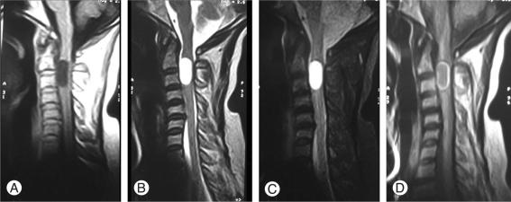
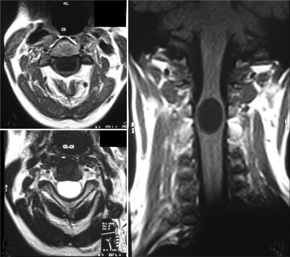
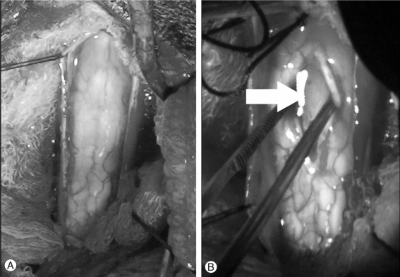
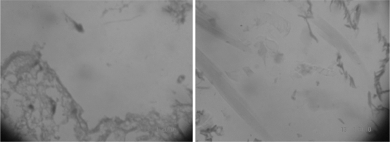
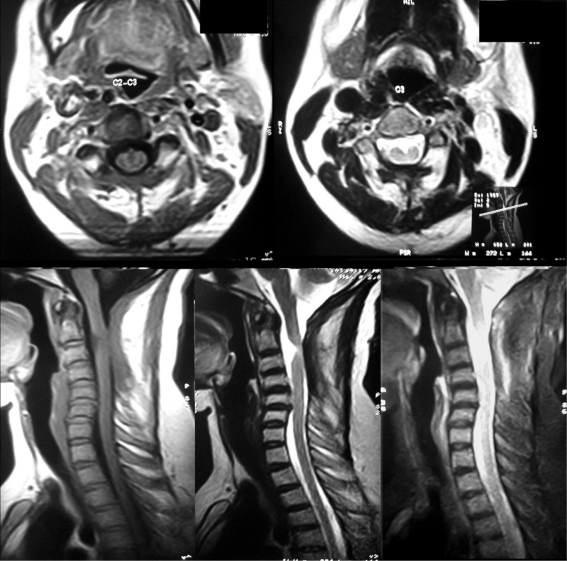
Intramedullary spinal epidermoid cysts are benign ectopic embryological growths with reported incidence of less than 1% of intramedullary tumors. In this case we report an unusual cervical intramedullary epidermid with liquid contents. A 40-year-old patient presented with progressive weakness of all four limbs of four months duration, bowel and bladder disturbances of two days duration, pain and paresthesias in all four limbs. Magnetic resonance imaging (MRI) revealed a well defined intramedullary lesion extending from C2-C3 level with widening of the cord. The lesion was hypointense on T1W images, hyperintense on T2W and fluid attenuation and inversion recovery images with thin rim of enhancement after contrast administration. Histopathological examination of the excised specimen revealed epidermal lining and keratinous material features of an epidermoid cyst. As in present case, rarely epidermoid cyst can have clear contents, and an MRI finding can closely mimic the features of arachnoid cyst, findings not classical and is different than described in literature.
Keywords: Epidermal cyst; Intramedullary epidermoid; Magnetic resonance imaging; Spinal cord neoplasms.
Figures
References
-
- Ogden AT, Khandji AG, McCormick PC, Kaiser MG. Intramedullary inclusion cysts of the cervicothoracic junction: report of two cases in adults and review of the literature. J Neurosurg Spine. 2007;7:236–242. - PubMed
-
- Tekkök IH. Intramedullary epidermoid cysts. J Neurosurg Spine. 2008;8:202–203. - PubMed
-
- Kukreja K, Manzano G, Ragheb J, Medina LS. Differentiation between pediatric spinal arachnoid and epidermoid-dermoid cysts: is diffusion-weighted MRI useful. Pediatr Radiol. 2007;37:556–560. - PubMed
-
- Bloomer CW, Ackerman A, Bhatia RG. Imaging for spine tumors and new applications. Top Magn Reson Imaging. 2006;17:69–87. - PubMed
-
- Teksam M, Casey SO, Michel E, Benson M, Truwit CL. Intraspinal epidermoid cyst: diffusion-weighted MRI. Neuroradiology. 2001;43:572–574. - PubMed
LinkOut - more resources
Full Text Sources
Research Materials
Miscellaneous
