Posterior reversible encephalopathy syndrome in a case of postoperative spinal extradural haematoma: case report and review of literature
- PMID: 21386948
- PMCID: PMC3047900
- DOI: 10.4184/asj.2011.5.1.64
Posterior reversible encephalopathy syndrome in a case of postoperative spinal extradural haematoma: case report and review of literature
Abstract
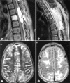
A 14-year-old girl presented with progressive paraparesis and paresthesia of one-year duration. Magnetic resonance imaging revealed a T6 vertebral hemangioma with epidural compression on the spinal cord. Following angiography and embolization, she underwent dorsal laminectomy and excision of the soft tissue component compressing the cord. In the postoperative period she had rapid worsening of lower limb power and imaging demonstrated an epidural haematoma at the operative site. The patient was taken up for urgent re-exploration and evacuation of haematoma. Postoperatively the patient complained of visual failure, headache and had multiple episodes of seizures. An magnetic resonance imaging brain showed characteristic features of posterior reversible encephalopathy syndrome (PRES) and the patient improved gradually after control of hypertension. This is the first documented case of PRES following spinal cord compression in a patient without any known risk factors. We postulate the possible mechanism involved in its pathogenesis.
Keywords: Hematoma; Magnetic resonance imaging; Posterior reversible encephalopathy syndrome; epidural; spinal.
Figures

References
-
- Hinchey J, Chaves C, Appignani B, et al. A reversible posterior leukoencephalopathy syndrome. N Engl J Med. 1996;334:494–500. - PubMed
-
- Gümüs H, Per H, Kumandas S, Yikilmaz A. Reversible posterior leukoencephalopathy syndrome in childhood: report of nine cases and review of the literature. Neurol Sci. 2010;31:125–131. - PubMed
-
- Lamy C, Oppenheim C, Méder JF, Mas JL. Neuroimaging in posterior reversible encephalopathy syndrome. J Neuroimaging. 2004;14:89–96. - PubMed
-
- Schwartz RB, Feske SK, Polak JF, et al. Preeclampsia-eclampsia: clinical and neuroradiographic correlates and insights into the pathogenesis of hypertensive encephalopathy. Radiology. 2000;217:371–376. - PubMed
LinkOut - more resources
Full Text Sources

