. 2011 Feb 28;6(2):e17027.
doi: 10.1371/journal.pone.0017027.

Acinetobacter baumannii secretes cytotoxic outer membrane protein A via outer membrane vesicles

Affiliations

Acinetobacter baumannii secretes cytotoxic outer membrane protein A via outer membrane vesicles

Jong Sook Jin et al. PLoS One. .

Abstract

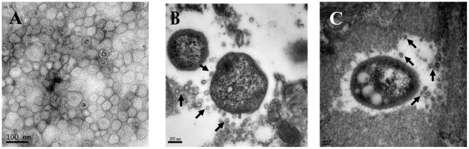
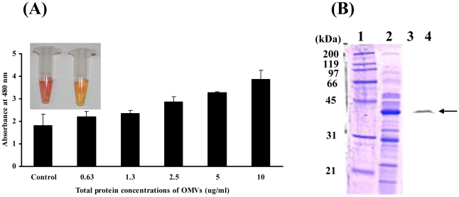
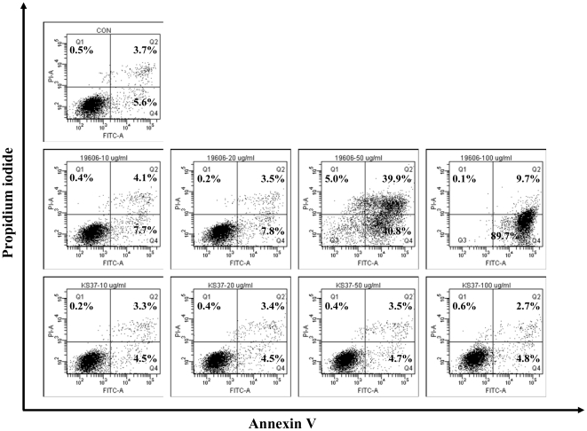
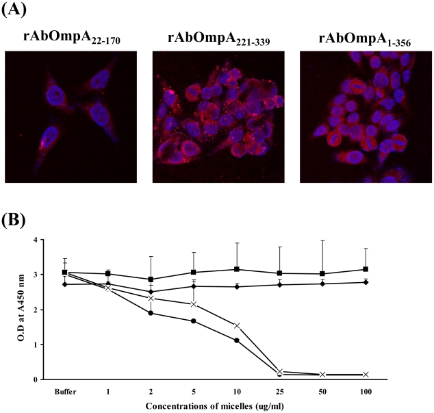
Acinetobacter baumannii is an important nosocomial pathogen that causes a high morbidity and mortality rate in infected patients, but pathogenic mechanisms of this microorganism regarding the secretion and delivery of virulence factors to host cells have not been characterized. Gram-negative bacteria naturally secrete outer membrane vesicles (OMVs) that play a role in the delivery of virulence factors to host cells. A. baumannii has been shown to secrete OMVs when cultured in vitro, but the role of OMVs in A. baumannii pathogenesis is not well elucidated. In the present study, we evaluated the secretion and delivery of virulence factors of A. baumannii to host cells via the OMVs and assessed the cytotoxic activity of outer membrane protein A (AbOmpA) packaged in the OMVs. A. baumannii ATCC 19606(T) secreted OMVs during in vivo infection as well as in vitro cultures. Potential virulence factors, including AbOmpA and tissue-degrading enzymes, were associated with A. baumannii OMVs. A. baumannii OMVs interacted with lipid rafts in the plasma membranes and then delivered virulence factors to host cells. The OMVs from A. baumannii ATCC 19606(T) induced apoptosis of host cells, whereas this effect was not detected in the OMVs from the ΔompA mutant, thereby reflecting AbOmpA-dependent host cell death. The N-terminal region of AbOmpA(22-170) was responsible for host cell death. In conclusion, the OMV-mediated delivery of virulence factors to host cells may well contribute to pathogenesis during A. baumannii infection.


Conflict of interest statement

Competing Interests: The authors have declared that no competing interests exist.

Figures

Figure 1
Figure 1. Secretion of OMVs from A. baumannii during in vitro culture and in vivo infection.
(A) Transmission electron micrograph of OMVs prepared from A. baumannii ATCC 19606T cultured in LB broth for 24 h. (B and C) Secretion of OMVs from A. baumannii ATCC 19606T in a murine pneumonia model. Mice were infected with 1×10⁷ CFU of bacteria intratracheally and sacrificed 48 h after bacterial injection. Arrows indicate the OMVs secreted from A. baumannii.
Figure 2
Figure 2. A. baumannii OMVs contain biologically active proteins.
(A) A. baumannii OMVs were incubated with 100 mM nitrocefin in PBS at 30°C for 30 min. Hydrolysis of nitrocefin was monitored at 480 nm using a UV spectrophotometer. Inlet figure: left, brown colorization of nitrocefin in A. baumannii OMVs; right, negative control of nitrocefin in PBS. (B) SDS-PAGE of proteins packaged in the OMVs from A. baumannii ATCC 19606T (lanes 1 and 2) and its Western blot analysis (lanes 3 and 4). The samples were immunoblotted with a rabbit anti-AbOmpA immune serum. Lanes 1 and 3, molecular weight maker; 2 and 4, OMV fraction. Arrows indicate AbOmpA.
Figure 3
Figure 3. A. baumannii OMVs deliver virulence factor AbOmpA into host cells.
(A) HeLa and HEp-2 cells were treated with A. baumannii OMVs (5 µg/ml of protein concentrations) for 12 h. The cells were fixed, permeabilized, and stained with anti-rabbit AbOmpA antibody, followed by Alexa Fluor® 568-conjugated rabbit IgG (red). DAPI was used to stain the nuclei (blue). Magnification: ×400. The U937 cells were treated with 5 µg/ml of A. baumannii OMVs for 4 h and then stained with anti-rabbit AbOmpA antibody, followed by Alexa Fluor® 488-conjugated rabbit IgG (green). Magnification: ×1,260. (B) Western blot analysis of cell lysates. The differentiated U937 cells were treated with A. baumannii OMVs (20 µg/ml of protein concentrations) for the indicated times. Cell lysates were separated on 12% SDS-PAGE, transferred to membranes, and immunoblotted with a rabbit anti-AbOmpA immune serum and β-actin antibody.
Figure 4
Figure 4. A. baumannii OMVs interact with plasma membrane of host cells through a cholesterol-rich membrane microdomain.
(A) HeLa cells were treated with OMVs (5 µg/ml of protein concentrations) for 4 h. (B) HeLa cells were preterated with 10 mM MβCD for 45 min and then treated with OMVs for 4 h. The cells were fixed, permeabilized, and stained with anti-rabbit AbOmpA antibody, followed by Alexa Fluor® 488-conjugated rabbit IgG (green). DAPI was used to stain the nuclei (blue). Magnification: ×630.
Figure 5
Figure 5. Flow cytometric analysis of cell death induced by the OMVs from A. baumannii ATCC 19606T and the ΔompA mutant.
The differentiated U937 cells were treated with various concentrations (0, 10, 20, 50, and 100 µg/ml) of OMVs and stained with Annexin V and PI. Upper panel, control cells without OMVs for 24 h. Middle panel, the cells were treated with OMVs from A. baumannii ATCC 19606T for 24 h. Lower panel, the cells were treated with OMVs from the ΔompA mutant for 24 h. Representative data from three independent experiments are shown. In the graph, cells in right upper and lower parts are apoptotic cells and cells in left upper part are necrotic cells.
Figure 6
Figure 6. Host cell entry and cytotoxicity of rAbOmpA fragments.
(A) HeLa cells were treated with different forms of micelles containing rAbOmpA fragments for 24 h. The cells were fixed, permeabilized, and stained with anti-rabbit AbOmpA antibody, followed by Alexa Fluor® 568-conjugated rabbit IgG (red). DAPI was used to stain the nuclei (blue). The subcellular localization of AbOmpA was observed by confocal microscopy. Magnification: ×400. (B) The differentiated U937 cells were treated with different forms of micelles containing rAbOmpA fragments for 24 h. Cell viability was determined by a WST-1 assay. Untreated control cells (⧫), rAbOmpA22-170 (•), rAbOmpA1-356 (X), and rAbOmpA221-339 (▪).

Similar articles

Cited by

References

    1. Dijkshoorn L, Nemec A, Seifert H. An increasing threat in hospitals: multidrug-resistant Acinetobacter baumannii. Nat Rev Microbiol. 2007;5:939–951. - PubMed
    1. Peleg AY, Seifert H, Paterson DL. Acinetobacter baumannii: emergence of a successful pathogen. Clin Microbiol Rev. 2008;21:538–582. - PMC - PubMed
    1. Gordon MC, Wareham DW. Multidrug-resistant Acinetobacter baumannii: mechanisms of virulence and resistance. Int J Antimicrob Agents. 2010;35:219–226. - PubMed
    1. Falagas ME, Bliziotis IA, Kasiakou SK, Samonis G, Thanassopoulou P, et al. Outcome of infections due to pandrug-resistant (PDR) Gram-negative bacteria. BMC Infect Dis. 2005;5:24–30. - PMC - PubMed
    1. Smith MG, Gianoulis TA, Pukatzki S, Mekalanos JJ, Ornston LN, et al. New insights into Acinetobacter baumannii pathogenesis revealed by high-density pyrosequencing and transposon mutagenesis. Genes Dev. 2007;21:601–614. - PMC - PubMed

Publication types

MeSH terms

Substances

LinkOut - more resources