. 2011 Feb 28;6(2):e17241.
doi: 10.1371/journal.pone.0017241.

Two phosphodiesterase genes, PDEL and PDEH, regulate development and pathogenicity by modulating intracellular cyclic AMP levels in Magnaporthe oryzae

Affiliations

Two phosphodiesterase genes, PDEL and PDEH, regulate development and pathogenicity by modulating intracellular cyclic AMP levels in Magnaporthe oryzae

Haifeng Zhang et al. PLoS One. .

Abstract

Cyclic AMP (cAMP) signaling plays an important role in regulating multiple cellular responses, such as growth, morphogenesis, and/or pathogenicity of eukaryotic organisms such as fungi. As a second messenger, cAMP is important in the activation of downstream effector molecules. The balance of intracellular cAMP levels depends on biosynthesis by adenylyl cyclases (ACs) and hydrolysis by cAMP phosphodiesterases (PDEases). The rice blast fungus Magnaporthe oryzae contains a high-affinity (PdeH/Pde2) and a low-affinity (PdeL/Pde1) PDEases, and a previous study showed that PdeH has a major role in asexual differentiation and pathogenicity. Here, we show that PdeL is required for asexual development and conidial morphology, and it also plays a minor role in regulating cAMP signaling. This is in contrast to PdeH whose mutation resulted in major defects in conidial morphology, cell wall integrity, and surface hydrophobicity, as well as a significant reduction in pathogenicity. Consistent with both PdeH and PdeL functioning in cAMP signaling, disruption of PDEH only partially rescued the mutant phenotype of ΔmagB and Δpka1. Further studies suggest that PdeH might function through a feedback mechanism to regulate the expression of pathogenicity factor Mpg1 during surface hydrophobicity and pathogenic development. Moreover, microarray data revealed new insights into the underlying cAMP regulatory mechanisms that may help to identify potential pathogenicity factors for the development of new disease management strategies.


Conflict of interest statement

Competing Interests: The authors have declared that no competing interests exist.

Figures

Figure 1
Figure 1. The ΔpdeL and ΔpdeH mutants have defects in conidial morphology and hyphal branching.
(A) Conidia of the wild type and mutants were observed under an epi-fluorescence microscope. Conidia were stained with 1 µg/ml Calcofluor white (CFW) for 5 min in dark. (B) Conidial size of the wild type and mutants. Values are the mean ±SD from 100 conidia for each strain, which were measured using a microscope ruler. Length is the distance from the base to apex of conidia. And width is the size of the longest septum. (C) Branching patterns of mycelia on complete media slides at day 2 after incubation. Frequent branching occurs at the terminal mycelia of ΔpdeH and ΔpdeHΔpdeL double mutants. Calcofluor staining of mycelia is used to show the distance of septa.
Figure 2
Figure 2. ΔpdeH mutants have a defect in cell-wall integrity.
Growth of wild type and mutant strains on complete media (CM) without 1 M sorbitol (top); growth of strains on CM with 1 M sorbitol (middle); growth of strains on straw decoction and corn media (SDC) without sorbitol (bottom). The ΔpdeH and ΔpdeH ΔpdeL mutants undergo progressive autolysis on CM in the absence of osmotic stabilization. Radial growth rates are identical to those of the wild-type strains.
Figure 3
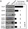
Figure 3. Detergent wettable phenotype of ΔpdeH and ΔpdeHΔpdeL mutants.
(A) Ten microliters of water or detergent solution containing 0.02% SDS and 5 mM EDTA were placed on the colony surfaces of the wild type and mutants strains and photographed after 5 min. (B) Expression analysis of MPG1 gene in wild type and mutant strains. The error bars indicate SD of three replicates. Asterisk indicates significant differences at P = 0.01.
Figure 4
Figure 4. Over-expression MPG1 in the ΔpdeH mutant partially restores the surface hydrophobicity and pathogenicity defects.
(A) Ten microliters of water or detergent solution containing 0.02% SDS and 5 mM EDTA were placed on the colony surfaces of wild type, mutants and Mpg1 over-expression strains and photographed after 5 min. Expression analysis of MPG1 gene in wild type and mutants and MPG1 over-expression strains. The error bars indicate SD of three replicates. Asterisk indicates significant differences at P = 0.01. (B) Spraying assay. Five milliliters of conidial suspension (5×10⁴ spores/ml) of each strain were sprayed on rice seedlings. Diseased leaves were photographed 7 days after inoculation.
Figure 5
Figure 5. PdeL and PdeH regulate intracellular cAMP levels during pathogenesis.
Loss of PDEL and PDEH leads to increased accumulation of cAMP levels. Bar chart showing quantification of intracellular cAMPs in the mycelia of the indicated strains cultured for 2 days in complete medium. Two biological repetitions with three replicates were assayed. The error bars represent SD of three replicates.
Figure 6
Figure 6. Appressorium formation assays.
(A) Conidia of each strain were incubated on hydrophobic surfaces for 24 hours (up panel); hyphal plugs of each strain were incubated on hydrophobic surfaces for 48 hours (bottom panel). (B) Conidia of each strain were incubated on hydrophilic surfaces for 24 hours.
Figure 7
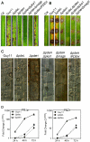
Figure 7. The loss of PDEH leads to reduced pathogenicity and induction of strong plant defense responses.
(A) Spraying assay. Five milliliters of conidial suspension (5×10⁴ spores/ml) of each strain were sprayed on rice seedlings. Diseased leaves were photographed 7 days after inoculation. (B) Detached leaf assay. The hyphal plugs of each strain were placed onto the upside of detached rice seedling leaves. Diseased leaves were photographed 5 days after inoculation. (C) Observation of infectious growth. Excised rice sheath from 4-week-old rice seedlings was inoculated with conidial suspension (5×10⁴ spores/ml of each strain). Infectious growth was observed 48 hours after inoculation. (D) The expression of rice pathogenesis-related (PR) genes over time after inoculation. The transcriptional expression of PR1a and PBZ1 in the infected rice was analyzed using quantitative RT-PCR. The graph was generated with three replicates in a representative data set, and similar results were obtained in another independent biological repetition. The error bars indicate SD of three replicates.
Figure 8
Figure 8. PdeL and PdeH are related to the activity of extracellular laccases.
(A) Laccase activity was tested on CM agar medium containing 0.2 mM ABTS at final concentration. Discoloration was observed on day 2. (B) Laccase activity measured by ABTS oxidizing test (see Materials and Methods for details). (C) Quantitative RT-PCR analysis of two laccase genes in wild type and mutants. Expression data were normalized using the ACTIN gene. Error bars represent standard deviation.
Figure 9
Figure 9. Functional categorization of the consensus genes.
(A) Expression profiles were combined and showing PdeL- and PdeH-dependent. (B) Up-regulation and down-regulation of more than two-fold change genes were grouped according to their putative function (see Supplemental data for details).
Figure 10
Figure 10. PTH11 gene expression in ΔpdeH and ΔpdeL mutants.
RNA was extracted from mycelia cultured in liquid CM medium for 2 days. ACTIN was used for normalization, and the values were calculated by 2-ddCT methods with quantitative RT-PCR data. Values represent mean ±SD from two independent experiments with three replicates each. Asterisk indicates significant differences at P = 0.01.
Figure 11
Figure 11. Assessment for appressorium formation and pathogenicity of PdeH targeted gene deletion mutants.
Gene expression was assessed according to description in text. Appressorium formation and pathogenicity assessment were also performed as described in text.

Similar articles

Cited by

References

    1. Daniel PB, Walker WH, Habener JF. Cyclic AMP signaling and gene regulation. Annu Rev Nutr. 1998;18:353–83. - PubMed
    1. Charbonneau H, Beier N, Walsh KA, Beavo JA. Identification of a conserved domain among cyclic nucleotide phosphodiesterases from diverse species. Proc Natl Acad Sci USA. 1986;83:9308–9312. - PMC - PubMed
    1. D’Angelo MA, Sanguineti S, Reece JM, Birnbaumer L, Torres HN, et al. Identification, characterization and subcellular localization of TcPDE1, a novel cAMP-specific phosphodiesterase from Trypanosoma cruzi. Biochem J. 2004;378:63–72. - PMC - PubMed
    1. De Voti JSG, Beach D, McLeod M. Interaction between ran1+ protein kinase and cAMP dependent protein kinase as negative regulators of fission yeast meiosis. EMBO J. 1991;10:3759–3768. - PMC - PubMed
    1. Dunlap PV, Callahan SM. Characterization of a periplasmic 3_:5_-cyclic nucleotide phosphodiesterase gene, cdpP, from the marine symbiotic bacterium Vibrio fischeri. J Bacteriol. 1993;175:4615–4624. - PMC - PubMed

Publication types

MeSH terms