. 2011 Feb 28;6(2):e17320.
doi: 10.1371/journal.pone.0017320.

Downregulation of VRK1 by p53 in response to DNA damage is mediated by the autophagic pathway

Affiliations

Downregulation of VRK1 by p53 in response to DNA damage is mediated by the autophagic pathway

Alberto Valbuena et al. PLoS One. .

Abstract

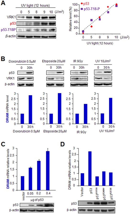
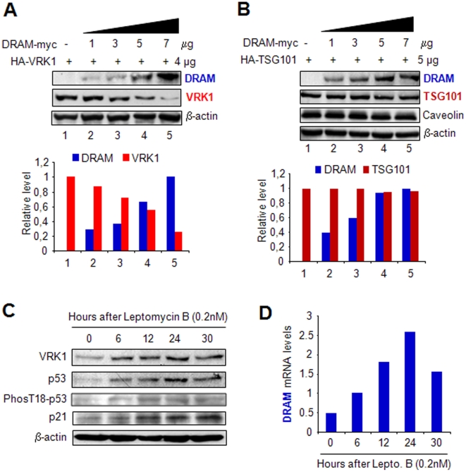
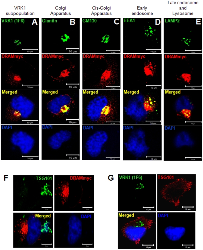
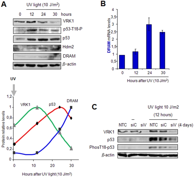
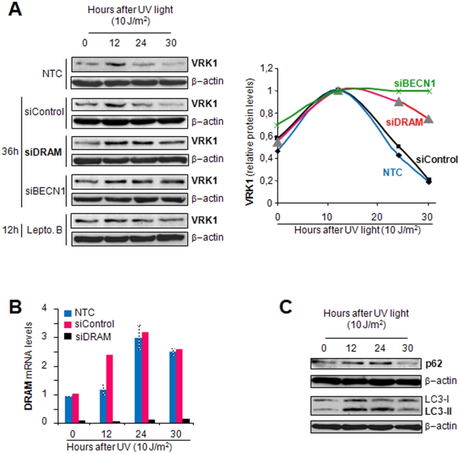
Human VRK1 induces a stabilization and accumulation of p53 by specific phosphorylation in Thr18. This p53 accumulation is reversed by its downregulation mediated by Hdm2, requiring a dephosphorylated p53 and therefore also needs the removal of VRK1 as stabilizer. This process requires export of VRK1 to the cytosol and is inhibited by leptomycin B. We have identified that downregulation of VRK1 protein levels requires DRAM expression, a p53-induced gene. DRAM is located in the endosomal-lysosomal compartment. Induction of DNA damage by UV, IR, etoposide and doxorubicin stabilizes p53 and induces DRAM expression, followed by VRK1 downregulation and a reduction in p53 Thr18 phosphorylation. DRAM expression is induced by wild-type p53, but not by common human p53 mutants, R175H, R248W and R273H. Overexpression of DRAM induces VRK1 downregulation and the opposite effect was observed by its knockdown. LC3 and p62 were also downregulated, like VRK1, in response to UV-induced DNA damage. The implication of the autophagic pathway was confirmed by its requirement for Beclin1. We propose a model with a double regulatory loop in response to DNA damage, the accumulated p53 is removed by induction of Hdm2 and degradation in the proteasome, and the p53-stabilizer VRK1 is eliminated by the induction of DRAM that leads to its lysosomal degradation in the autophagic pathway, and thus permitting p53 degradation by Hdm2. This VRK1 downregulation is necessary to modulate the block in cell cycle progression induced by p53 as part of its DNA damage response.


Conflict of interest statement

Competing Interests: The authors have declared that no competing interests exist.

Figures

Figure 1
Figure 1. Effect of p53 on the transcription of endogenous DRAM gene.
(A) Determination of the optimal dose of UV light that induces p53 stabilization and its phosphorylation in Thr18 in the WS1 cell line. To the right is shown the quantification of p53 and p53 phosphorylated in Thr18 as a function of the UV dose. (B) Different types of DNA damage induce endogenous p53 accumulation, and VRK1 downregulation in WS1 fibroblasts (p53+/+) determined by western blot (top). DNA damage also induces DRAM accumulation detected by qRT-PCR in human WS1 fibroblasts. The DNA damage agents used were doxorubicin, etoposide, ionizing radiation and UV-C light (254 nm). (C) H1299 (p53−/−) cells transfected with increasing amounts of plasmid pCB6+p53 and expression of DRAM was determined by qRT-PCR. Values are the mean of three experiments with standard deviation. Same amount of DNA was used in all transfections that were completed with empty vector as necessary. (D) H1299 (p53−/−) were transfected with the indicated plasmids pCB6+p53 (wt), pCMV-p53R175H, pCMV-p53R248W and pCMV-p53R273H, and the effect on the expression of endogenous DRAM gene expression was determined by qRT-PCR. In the immunoblots (IB) at the bottom is shown the correct expression of the different p53 proteins, wild-type or mutants.
Figure 2
Figure 2. Dose dependent effect of DRAM on VRK1, and TSG101 protein levels.
The variation in levels of VRK1 (A), and the control endosomal protein TSG101 implicated endocytic recycling of plasma membrane receptors and caveolin (B) were determined in response to increasing levels of DRAM. H1299 (p53−/−) cells were transfected with a fixed amount of VRK1 and TSG101 plasmids, and varying concentrations of DRAM. Plasmid pCDNA3-DRAM-Myc-His , plasmid pCEFL-HA-VRK1 , and plasmid pHA-TSG101 were used. To the bottom is shown the quantification of corresponding immunoblots in the linear response range. VRK1 and TSG101 proteins were detected with an anti HA antibody. DRAM was detected with an anti myc antibody. C. The block of nuclear export by leptomycin B in WS1 cell line prevented degradation of VRK1 even though there is an accumulation p53, which is also phosphorylated in Thr18 and induces DRAM gene expression. D. Accumulation of p53 by leptomycin B induces DRAM gene expression in WS1 cells as determined by quantitative qRT-PCR.
Figure 3
Figure 3. Subcellular localization of DRAM.
(A–E) Colocalization of DRAM in different Golgi-endosome-lysosome compartments. (F) TSG101 and DRAM do not colocalize in endoplasmic vesicles. (G) TSG101 and VRK1 colocalize in different locations in the absence of DRAM. In these experiments, H1299 cells were transfected with plasmid pCDNA3-DRAM-Myc-His and plated on 10-cm2 dishes (5×10⁵) containing 1-cm-diameter sterile glass coverslips. The coverslips were stained twenty-four hours after DRAM transfection with specific antibodies for the endogenous proteins: VRK1 (1F6), giantin, GM130, EEA1 and LAMP2. DRAM was detected with an anti-myc epitope antibody. TSG101 was detected with a polyclonal antibody.
Figure 4
Figure 4. Sequential changes in VRK1, p53 and DRAM protein levels induced by UV in WS1 human fibroblasts.
(A) Levels of the three proteins VRK1, p53 and DRAM after treating human fibroblasts WS1 with UV light. The relative level of each protein was quantified and represented in the graph at the bottom. (B) Quantification of DRAM RNA levels by qRT-PCR at different time points following treatment with UV light. Mean of three independents experiments with standard deviation. (C). Knockdown of VRK1 (siV), but not controls (siC and NTC), prevented the accumulation of p53 and its phosphorylation in Thr18 in response to UV irradiation. Knock-down siRNA transfections were performed 96 hours before the start of UV treatment. Cell lysates were prepared 12 hours after irradiation.
Figure 5
Figure 5. Knock-down of DRAM and Beclin-1 (BECN1), and addition of leptomycin B prevented downregulation of VRK1 induced by UV light.
(A) Human fibroblast WS1 cells were transfected with siControl, siDRAM-01, siBECN1-smart pool, or treated with leptomycin B. After that, these cells were irradiated with UV-C light and the protein levels determined at different time points. The changes in levels of VRK1 protein were detected with the 1B5 mAb. The quantification of the blots is shown in the graph at the right. (B). The effectiveness of the DRAM knock-down was determined by qRT-PCR and the result shown in the bar graph at the bottom, and siBECN1 by western blot (Fig. S2). Knock-down siRNA transfections were performed 48 hours, and addition of leptomycin B was 12 hours, before the start of UV treatment. (C) P62/SQSTM1 and LC3B are proteins degraded by autophagy. P62/SQSTM1 and LC3B proteins are also degraded in response to UV light, following a transient accumulation in autophagosomes after induction of damage , .
Figure 6
Figure 6. Model of the autoregulatory VRK1-p53-Hdm2-DRAM loop.
Several types of DNA damage mechanisms can induce VRK1, stabilizing and activating p53-dependent transcription (black line). Among the p53-induced genes, Hdm2 promotes p53 degradation via ubiquitylation (blue line), and DRAM induces VRK1 degradation in the lysosome (red line). VRK1 and DRAM are in the same late endosomal vesicle that fuses to lysosomes, but do not interact directly. PP: unknown phosphatase.

Similar articles

Cited by

References

    1. Sengupta S, Harris CC. p53: traffic cop at the crossroads of DNA repair and recombination. Nat Rev Mol Cell Biol. 2005;6:44–55. - PubMed
    1. Horn HF, Vousden KH. Coping with stress: multiple ways to activate p53. Oncogene. 2007;26:1306–1316. - PubMed
    1. Baehrecke EH. Autophagy: dual roles in life and death? Nat Rev Mol Cell Biol. 2005;6:505–510. - PubMed
    1. Brech A, Ahlquist T, Lothe RA, Stenmark H. Autophagy in tumour suppression and promotion. Mol Oncol. 2009;3:366–375. - PMC - PubMed
    1. Saito S, Yamaguchi H, Higashimoto Y, Chao C, Xu Y, et al. Phosphorylation Site Interdependence of Human p53 Post-translational Modifications in Response to Stress. J Biol Chem. 2003;278:37536–37544. - PubMed

Publication types

MeSH terms