. 2011 Feb 28;6(2):e17385.
doi: 10.1371/journal.pone.0017385.

Relationship between neural alteration and perineural invasion in pancreatic cancer patients with hyperglycemia

Affiliations

Relationship between neural alteration and perineural invasion in pancreatic cancer patients with hyperglycemia

Junhui Li et al. PLoS One. .

Abstract

Background: Patients with higher levels of fasting serum glucose have higher death rates from pancreatic cancer compared to patients with lower levels of fasting serum glucose. However, the reasons have not been studied. The goal of the current study was to examine the neural alterations in pancreatic cancer patients with hyperglycemia and to identify the relationship between the neural alterations and perineural invasion.

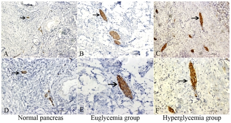
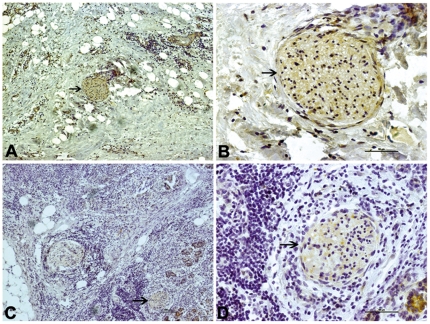
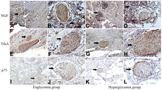
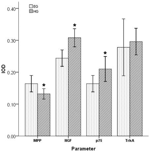
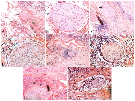
Methodology/principal findings: The clinical and pathological features of 61 formalin-fixed pancreatic cancer specimens and 10 normal pancreases as controls were analyzed. Furthermore, the expression of Protein Gene Product 9.5 (PGP9.5), Myelin P0 protein (MPP), NGF, TrkA, and p75 were examined by immunohistochemistry. The median number of nerves, the median area of neural tissue, and the median nerve diameter per 10 mm(2) were larger in the hyperglycemia group than those in the euglycemia group (p = 0.007, p = 0.009, and p = 0.004, respectively). The integrated optical density (IOD) of MPP staining was lower in the hyperglycemia group than those in the euglycemia group (p = 0.019), while the expression levels of NGF and p75 were higher in the hyperglycemia group than those in the euglycemia group (p = 0.002, and p = 0.026, respectively). The nerve bundle invasion of pancreatic cancer was more frequent in the hyperglycemia group than in the euglycemia group (p = 0.000).

Conclusions/significance: Nerve damage and regeneration occur simultaneously in the tumor microenvironment of pancreatic cancer patients with hyperglycemia; the simultaneous occurrence may aggravate the process of perineural invasion. The abnormal expression of NGF and p75 may also be involved in this process and subsequently lead to a lower rate of curative surgery.


Conflict of interest statement

Competing Interests: The authors have declared that no competing interests exist.

Figures

Figure 1
Figure 1. Nerve tissue immunostaining in different group.
PGP 9.5 immunostaining in the normal pancreas (A, D, arrowhead), pancreatic cancer in euglycemia group (B, E, arrowhead), and hyperglycemia group (C, F, arrowhead). A, B, C show an original magnification of 100×, and D, E, F an original magnification of 200×. In the hyperglycemia group, comparable increases in the number, area, and diameter of nerve tissues were present. Figure arrowheads indicate the immunostainings.
Figure 2
Figure 2. Myelin sheath immunostaining in different group.
MPP immunostaining of pancreatic cancer tissue in the euglycemia group (A, B, arrowhead) and hyperglycemia group (C, D, arrowhead). The left panel shows an original magnification of 100×, and the right panel an original magnification of 400×. The frequency of moderate to strong MPP staining of nerves was significantly lower in the hyperglycemia group than in the euglycemia group. Figure arrowheads indicate the immunostainings.
Figure 3
Figure 3. NGF, TrkA, and p75 immunostaining of pancreatic cancer tissue in the euglycemia group and hyperglycemia group.
A, C, E, G, I, K show an original magnification of 100×, and B, D, F, H, J, L an original magnification of 400×. The frequency of moderate to strong NGF staining of cancer cells was significantly higher in the hyperglycemia group than that in the euglycemia group (arrowhead). The NGF stain of nerves was not significantly different between the two groups. The moderate to strong TrkA stainings were also present in the two groups (arrowhead) but with no significant difference. The frequency of moderate to strong p75 staining of nerves (arrowhead) was significantly higher in the hyperglycemia group than that in the euglycemia group. Figure arrowheads indicate the immunostainings.
Figure 4
Figure 4. The comparation of four parameters in different group.
Sixty-one patients who received a radical curative pancreatic operations at the First Affiliated Hospital, Xi'an Jiaotong University with a pathologic diagnose were investigated, which included 45 specimen in euglycemia group and 16 specimen in hyperglycemia group. The MPP, NGF, TrkA and p75 immunostainings were analyzed by Image-Pro Plus software. The integrated optical density (IOD) of MPP staining was lower in the hyperglycemia group than that in the euglycemia group (p = 0.019). The IOD of NGF and p75 stainings were higher in the hyperglycemia group than that in the euglycemia group (p = 0.002, p = 0.026). The IOD of TrkA staining was not significantly different between the two groups.
Figure 5
Figure 5. The classification of PNI in different group.
Ne0–no perineural invasion; Ne1–neurium invasion; Ne2–perineural space invasion; Ne3–nerve bundle invasion. The nerve structures (A, B) and representative stages of the invaded neural structure for classification (C–H). (A) A nerve bundle surrounded by the continuous layered epithelioid sheets of the perineurium (arrowhead). (B) The perineural space, an internal space between the nerve bundle and perineurium (arrowhead). (C) Neurium invasion of cancer cells in the outer perineural space and attached to the perineurium (arrowhead). (D) Perineurial cells of perineurium between the nerve bundle and cancer cells (arrowhead). (E) Perineural space invasion of cancer cells in the perineural space (arrowhead). (F) Cancer cells invading the perineural space and compressing the nerve bundle smoothly. The border between cancer cells and nerve bundles do not contain perineurial cells or perineurium (arrowhead). (G) Nerve bundle invasion by cancer cells. Cancer cells irregularly compress the nerve bundle, clearly invading between nerve fibers (arrowhead). (H) Invasive cancer cells clearly seen between nerve fibers (arrowhead). A, C, E, G show an original magnification of 100×, and B, D, F, H an original magnification of 400×. Figure arrowheads indicate the nerves.

References

    1. Hidalgo M. Pancreatic Cancer. N Engl J Med. 2010;362:1605–1617. - PubMed
    1. Dai H, Li R, Wheeler T, Ozen M, Ittmann M, et al. Enhanced survival in perineural invasion of pancreatic cancer: an in vitro approach. Hum pathol. 2007;38:299–307. - PubMed
    1. Nakao A, Harada A, Nonami T, Kaneko T, Takagi H. Clinical significance of carcinoma invasion of the extrapancreatic nerve plexus in pancreatic cancer. Pancreas. 1996;12:361–357. - PubMed
    1. Matsuda M, Nimura Y. Perineural invasion of pancreas head carcinoma. Nippon Geka Gakkai Zasshi. 1983;84:719–728. - PubMed
    1. Kayahara M, Nakagawara H, Kitagawa H, Ohta T. The Nature of Neural Invasion by Pancreatic Cancer. Pancreas. 2007;35:218–223. - PubMed

Publication types

MeSH terms