

Fucans, but not fucomannoglucuronans, determine the biological activities of sulfated polysaccharides from Laminaria saccharina brown seaweed
- PMID: 21387013
- PMCID: PMC3046160
- DOI: 10.1371/journal.pone.0017283
Fucans, but not fucomannoglucuronans, determine the biological activities of sulfated polysaccharides from Laminaria saccharina brown seaweed
Abstract
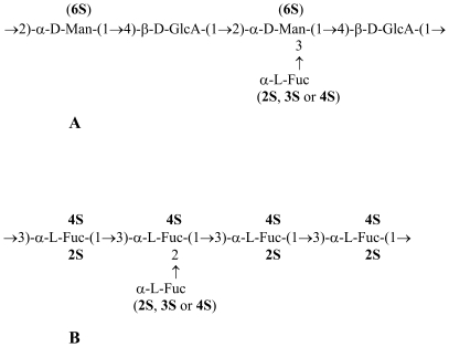
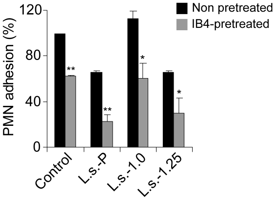
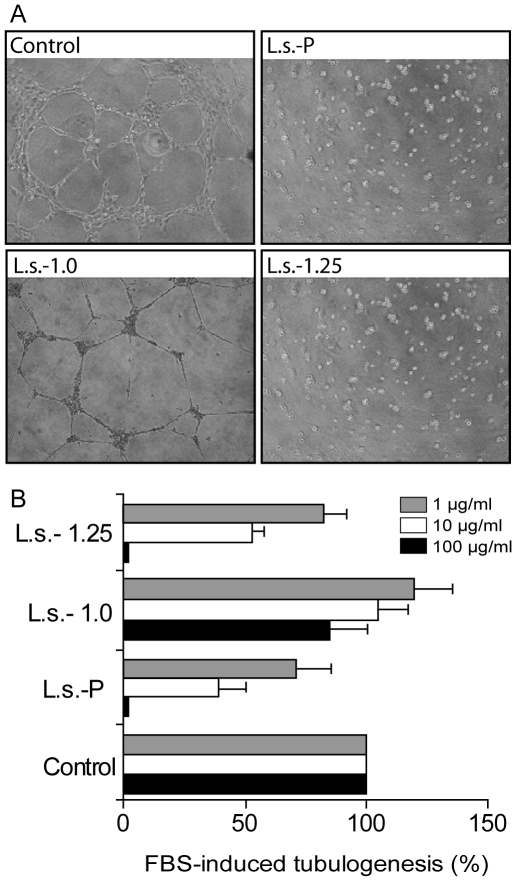
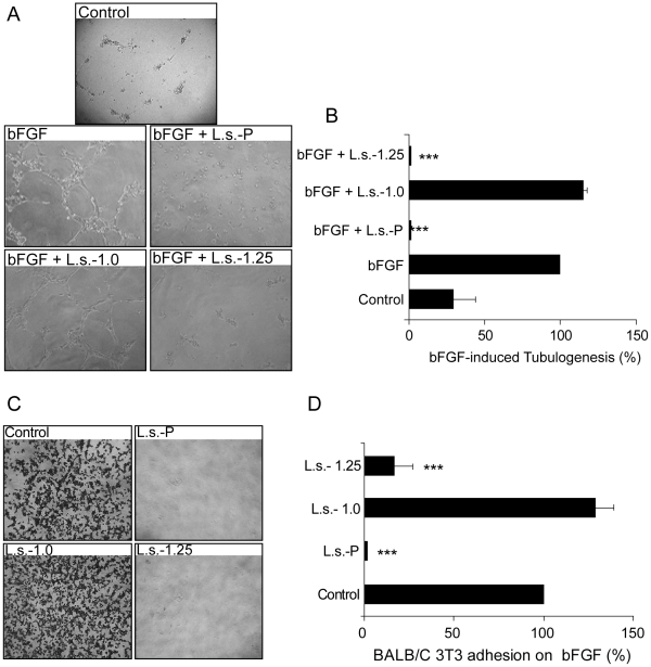
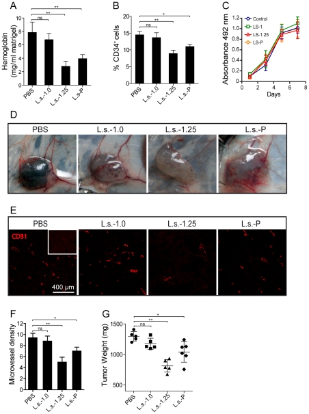
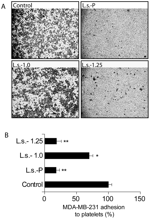
Sulfated polysaccharides from Laminaria saccharina (new name: Saccharina latissima) brown seaweed show promising activity for the treatment of inflammation, thrombosis, and cancer; yet the molecular mechanisms underlying these properties remain poorly understood. The aim of this work was to characterize, using in vitro and in vivo strategies, the anti-inflammatory, anti-coagulant, anti-angiogenic, and anti-tumor activities of two main sulfated polysaccharide fractions obtained from L. saccharina: a) L.s.-1.0 fraction mainly consisting of O-sulfated mannoglucuronofucans and b) L.s.-1.25 fraction mainly composed of sulfated fucans. Both fractions inhibited leukocyte recruitment in a model of inflammation in rats, although L.s.-1.25 appeared to be more active than L.s.-1.0. Also, these fractions inhibited neutrophil adhesion to platelets under flow. Only fraction L.s.-1.25, but not L.s.-1.0, displayed anticoagulant activity as measured by the activated partial thromboplastin time. Investigation of these fractions in angiogenesis settings revealed that only L.s.-1.25 strongly inhibited fetal bovine serum (FBS) induced in vitro tubulogenesis. This effect correlated with a reduction in plasminogen activator inhibitor-1 (PAI-1) levels in L.s.-1.25-treated endothelial cells. Furthermore, only parent sulfated polysaccharides from L. saccharina (L.s.-P) and its fraction L.s.-1.25 were powerful inhibitors of basic fibroblast growth factor (bFGF) induced pathways. Consistently, the L.s.-1.25 fraction as well as L.s.-P successfully interfered with fibroblast binding to human bFGF. The incorporation of L.s.-P or L.s.-1.25, but not L.s.-1.0 into Matrigel plugs containing melanoma cells induced a significant reduction in hemoglobin content as well in the frequency of tumor-associated blood vessels. Moreover, i.p. administrations of L.s.-1.25, as well as L.s.-P, but not L.s.-1.0, resulted in a significant reduction of tumor growth when inoculated into syngeneic mice. Finally, L.s.-1.25 markedly inhibited breast cancer cell adhesion to human platelet-coated surfaces. Thus, sulfated fucans are mainly responsible for the anti-inflammatory, anticoagulant, antiangiogenic, and antitumor activities of sulfated polysaccharides from L. saccharina brown seaweed.
Conflict of interest statement
Figures
Similar articles
-
A comparative study of the anti-inflammatory, anticoagulant, antiangiogenic, and antiadhesive activities of nine different fucoidans from brown seaweeds.Glycobiology. 2007 May;17(5):541-52. doi: 10.1093/glycob/cwm014. Epub 2007 Feb 12. Glycobiology. 2007. PMID: 17296677
-
Water-Soluble Saccharina latissima Polysaccharides and Relation of Their Structural Characteristics with In Vitro Immunostimulatory and Hypocholesterolemic Activities.Mar Drugs. 2023 Mar 16;21(3):183. doi: 10.3390/md21030183. Mar Drugs. 2023. PMID: 36976232 Free PMC article.
-
Sulfated galactofucan from Lobophora variegata: anticoagulant and anti-inflammatory properties.Biochemistry (Mosc). 2008 Sep;73(9):1018-24. doi: 10.1134/s0006297908090095. Biochemistry (Mosc). 2008. PMID: 18976219
-
Important determinants for fucoidan bioactivity: a critical review of structure-function relations and extraction methods for fucose-containing sulfated polysaccharides from brown seaweeds.Mar Drugs. 2011;9(10):2106-2130. doi: 10.3390/md9102106. Epub 2011 Oct 24. Mar Drugs. 2011. PMID: 22073012 Free PMC article. Review.
-
Use of sulfated fucans as anticoagulant and antithrombotic agents: future perspectives.Curr Pharm Des. 2004;10(9):967-81. doi: 10.2174/1381612043452730. Curr Pharm Des. 2004. PMID: 15078127 Review.
Cited by
-
Marine Compounds for Melanoma Treatment and Prevention.Int J Mol Sci. 2022 Sep 7;23(18):10284. doi: 10.3390/ijms231810284. Int J Mol Sci. 2022. PMID: 36142196 Free PMC article. Review.
-
Macroalgae-A Sustainable Source of Chemical Compounds with Biological Activities.Nutrients. 2020 Oct 11;12(10):3085. doi: 10.3390/nu12103085. Nutrients. 2020. PMID: 33050561 Free PMC article. Review.
-
A Comprehensive and Comparative Analysis of the Fucoidan Compositional Data Across the Phaeophyceae.Front Plant Sci. 2020 Nov 25;11:556312. doi: 10.3389/fpls.2020.556312. eCollection 2020. Front Plant Sci. 2020. PMID: 33324429 Free PMC article. Review.
-
Effects of oral administration of fucoidan extracted from Cladosiphon okamuranus on tumor growth and survival time in a tumor-bearing mouse model.Mar Drugs. 2012 Oct;10(10):2337-2348. doi: 10.3390/md10102337. Epub 2012 Oct 22. Mar Drugs. 2012. PMID: 23170088 Free PMC article.
-
Perspectives for the Use of Fucoidans in Clinical Oncology.Int J Mol Sci. 2022 Oct 5;23(19):11821. doi: 10.3390/ijms231911821. Int J Mol Sci. 2022. PMID: 36233121 Free PMC article. Review.
References
-
- Berteau O, Mulloy B. Sulfated fucans, fresh perspectives: structures, functions, and biological properties of sulfated fucans and an overview of enzymes active toward this class of polysaccharide. Glycobiology. 2003;13:29R–40R. - PubMed
-
- Cumashi A, Ushakova NA, Preobrazhenskaya ME, D'Incecco A, Piccoli A, et al. A comparative study of the anti-inflammatory, anticoagulant, antiangiogenic, and antiadhesive activities of nine different fucoidans from brown seaweeds. Glycobiology. 2007;17:541–552. - PubMed
-
- Kusaykin M, Bakunina I, Sova V, Ermakova S, Kuznetsova T, et al. Structure, biological activity, and enzymatic transformation of fucoidans from the brown seaweeds. Biotechnol J. 2008;3:904–915. - PubMed
-
- Ghosh T, Chattopadhyay K, Marschall M, Karmakar P, Mandal P, et al. Focus on antivirally active sulfated polysaccharides: from structure–activity analysis to clinical evaluation. Glycobiology. 2009;19:2–15. - PubMed
Publication types
MeSH terms
Substances
LinkOut - more resources
Full Text Sources
Other Literature Sources
Medical
Miscellaneous
