. 2011 Feb;69(2):303-11.
doi: 10.1002/ana.22297.

Investigations of caspr2, an autoantigen of encephalitis and neuromyotonia

Affiliations

Investigations of caspr2, an autoantigen of encephalitis and neuromyotonia

Eric Lancaster et al. Ann Neurol. 2011 Feb.

Abstract

Objective: To report clinical and immunological investigations of contactin-associated protein-like 2 (Caspr2), an autoantigen of encephalitis and peripheral nerve hyperexcitability (PNH) previously attributed to voltage-gated potassium channels (VGKC).

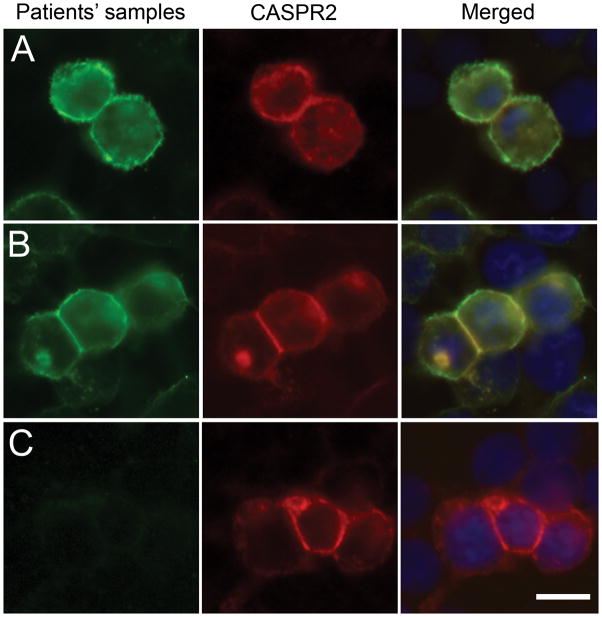
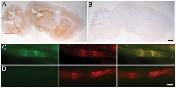
Methods: Clinical analysis was performed on patients with encephalitis, PNH, or both. Immunoprecipitation and mass spectrometry were used to identify the antigen and to develop an assay with Caspr2-expressing cells. Immunoabsorption with Caspr2 and comparative immunostaining of brain and peripheral nerve of wild-type and Caspr2-null mice were used to assess antibody specificity.

Results: Using Caspr2-expressing cells, antibodies were identified in 8 patients but not in 140 patients with several types of autoimmune or viral encephalitis, PNH, or mutations of the Caspr2-encoding gene. Patients' antibodies reacted with brain and peripheral nerve in a pattern that colocalized with Caspr2. This reactivity was abrogated after immunoabsorption with Caspr2 and was absent in tissues from Caspr2-null mice. Of the 8 patients with Caspr2 antibodies, 7 had encephalopathy or seizures, 5 neuropathy or PNH, and 1 isolated PNH. Three patients also had myasthenia gravis, bulbar weakness, or symptoms that initially suggested motor neuron disease. None of the patients had active cancer; 7 responded to immunotherapy and were healthy or only mildly disabled at last follow-up (median, 8 months; range, 6-84 months).

Interpretation: Caspr2 is an autoantigen of encephalitis and PNH previously attributed to VGKC antibodies. The occurrence of other autoantibodies may result in a complex syndrome that at presentation could be mistaken for a motor neuron disorder. Recognition of this disorder is important, because it responds to immunotherapy.


Conflict of interest statement

Conflicts of Interest

EL has received grant support from Talecris, a company which sells human immunoglobulin. A patent application for the use of Lgi1 antibody detection in patients’ CSF and sera has been filed by JD. No other authors have conflicts of interest.

Figures

Figure 1
Figure 1. Patients’ antibodies react with cells expressing Caspr2
HEK cells were transiently transfected to express human Caspr2, and labeled with the index patient’s CSF (A; green) or serum from a different patient (B; green) and a rabbit antibody to Caspr2 (A, B red), and counterstained with DAPI. Merged images (A, B yellow) demonstrate the overlap of patients’ antibody staining with Caspr2 expression. CSF (C) from a control patient did not react with cells expressing Caspr2. Scale bar: 20 μm
Figure 2
Figure 2. Immunoabsorption with Caspr2 abrogates the reactivity of the index patient’s antibodies with rodent brain and peripheral nerve
Top row: Rat brain sections were immunolabeled with the CSF of the index patient after immunoabsorption with sham transfected HEK cells (A) or HEK cells transfected to express Caspr2 (B); scale bar: 300 μm. Bottom rows: Teased mouse sciatic nerve fibers were immunolabeled with the CSF (green) of the index patient after immunoabsorption with sham transfected HEK cells (C) or HEK cells transfected to express Caspr2 (D); a rabbit antibody against Caspr2 (red) labels the endogenous Caspr2. Note that the reactivity of the patient’s CSF for both brain and teased fibers is abrogated when the CSF is preabsorbed with Caspr2. Scale bar: 10 μm.
Figure 3
Figure 3. Patients’ antibodies react with wild-type but not Caspr2-null mouse brains
The CSF (A) and serum (B) from 2 different patients with Caspr2 antibodies immunostain the hippocampus of a wild-type mouse in a pattern identical to that obtained with a rabbit polyclonal antibody against Caspr2 (C). This staining is not seen in sections from a Caspr2-null mouse (A–C, columns on the right). The pattern of staining with a CSF from a patient with limbic encephalitis associated with LGI1 antibodies is subtly different from Caspr2 and it is not affected using wild-type or Caspr2-null mice (D). The CSF of a control individual without Caspr2 or LGI1 antibodies does not label either sample (E). Scale bar: 100 μm
Figure 4
Figure 4. Patients’ antibodies label teased fibers from wild-type but not Caspr2-null nerves
The sera (green) of 2 patients with Caspr2 antibodies (A and B) immunolabel the juxtaparanodes of teased sciatic nerve fibers from wild-type mice in a pattern similar to that of a rabbit antibody against Caspr2 (red), but different than the paranodal staining seen with a mouse monoclonal against CASPR (blue). In contrast, neither these patients’ sera nor the rabbit antibody against Caspr2 label teased sciatic nerve fibers from Caspr2-null mice (columns on the right). Sera from 2 controls (C and D) do not label teased nerve fibers. Scale bar is 5 μm.

Similar articles

Cited by

References

    1. Lai M, Huijbers MG, Lancaster E, et al. Investigation of Lgi1 as the antigen of limbic encephalitis previously attributed to potassium channels. Lancet Neurol. 2010;9:776–785. - PMC - PubMed
    1. Buckley C, Oger J, Clover L, et al. Potassium channel antibodies in two patients with reversible limbic encephalitis. Ann Neurol. 2001;50:73–78. - PubMed
    1. Tan KM, Lennon VA, Klein CJ, et al. Clinical spectrum of voltage-gated potassium channel autoimmunity. Neurology. 2008;70:1883–1890. - PubMed
    1. Ances BM, Vitaliani R, Taylor RA, et al. Treatment-responsive limbic encephalitis identified by neuropil antibodies: MRI and PET correlates. Brain. 2005;128:1764–1777. - PMC - PubMed
    1. Bushara KO, Goebel SU, Shill H, et al. Gluten sensitivity in sporadic and hereditary cerebellar ataxia. Ann Neurol. 2001;49:540–543. - PubMed

Publication types

MeSH terms