

doi: 10.1021/ja110296z.
Epub 2011 Mar 9.
Computational design of the sequence and structure of a protein-binding peptide
Affiliations
- PMID: 21388199
- PMCID: PMC3081598
- DOI: 10.1021/ja110296z
Item in Clipboard
Computational design of the sequence and structure of a protein-binding peptide
J Am Chem Soc.
.
Abstract
The de novo design of protein-binding peptides is challenging because it requires the identification of both a sequence and a backbone conformation favorable for binding. We used a computational strategy that iterates between structure and sequence optimization to redesign the C-terminal portion of the RGS14 GoLoco motif peptide so that it adopts a new conformation when bound to Gα(i1). An X-ray crystal structure of the redesigned complex closely matches the computational model, with a backbone root-mean-square deviation of 1.1 Å.
Figures
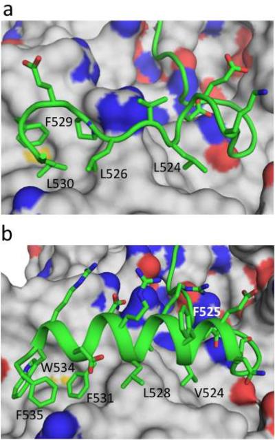
Redesigning the RGS14 GoLoco motif. (a) Crystal structure of the wildtype RGS14 GoLoco motif peptide bound to Gαi1. (b) Model of the redesigned GoLoco motif, GLhelix-4, bound to Gαi1. Both the wildtype GoLoco motif and GLhelix-4 are shown in cartoon representation with selected side chains displayed and labeled. Gαi1 is in surface representation with side-chain nitrogens colored blue and side-chain oxygens colored red.

Crystal Structure of GLhelix-4 bound to Gai1·GDP. The unbiased 2Fo-Fc electron density (blue mesh, contoured to σ=1.5, see Supplementary Methods), indicates a GoLoco motif peptide C-terminal α-helix (salmon) bound to the all-helical domain of Gαi1 (gray surface). The well-defined aromatic side chains Phe 525, Phe 531, and Trp 534 establish the correct helical orientation and register.

Model (light and dark green) and crystal structure (salmon and maroon) of GLhelix-4 bound to Gαi1. The structures were superimposed by minimizing the root mean square deviation between backbone heavy atoms in the all helical domain of Gαi1. The peptide side chains of Val 524 and Phe 531 pack on either side of Phe 108 from Gai1, as predicted. Only the orientation of Trp 534 and the absence of a definable residue 535 in the crystal structure deviate significantly from the predicted model.
References
Publication types
MeSH terms
Substances
Grants and funding
LinkOut - more resources
Full Text Sources
Other Literature Sources
