. 2011 Mar;175(3):266-81.
doi: 10.1667/RR2402.1. Epub 2010 Dec 20.

Blood-based detection of radiation exposure in humans based on novel phospho-Smc1 ELISA

Affiliations

Blood-based detection of radiation exposure in humans based on novel phospho-Smc1 ELISA

Richard G Ivey et al. Radiat Res. 2011 Mar.

Abstract

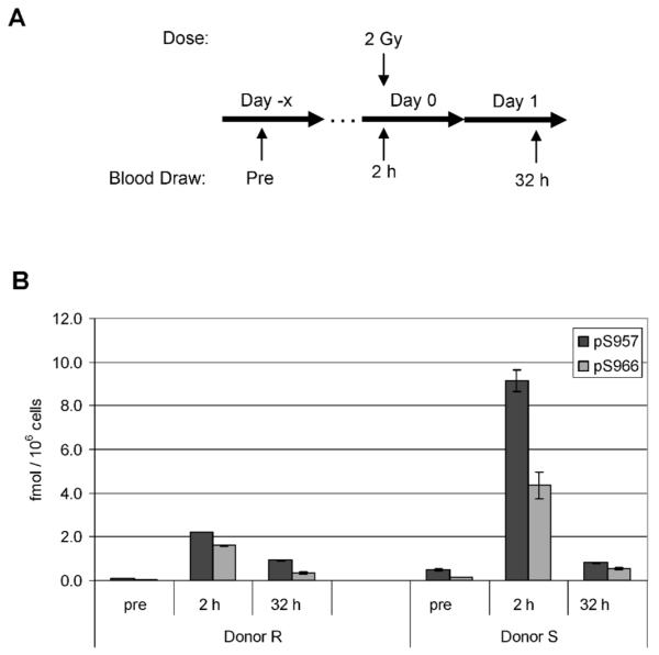
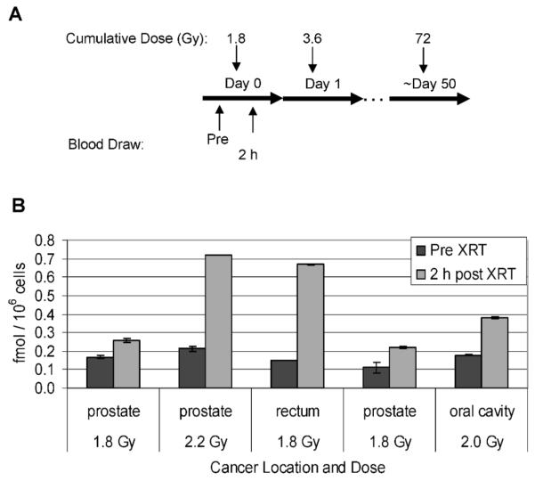
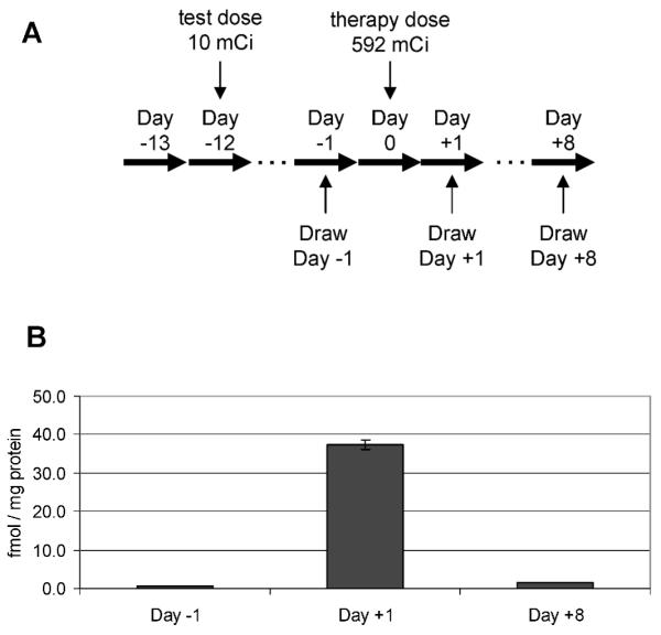
The structural maintenance of chromosome 1 (Smc1) protein is a member of the highly conserved cohesin complex and is involved in sister chromatid cohesion. In response to ionizing radiation, Smc1 is phosphorylated at two sites, Ser-957 and Ser-966, and these phosphorylation events are dependent on the ATM protein kinase. In this study, we describe the generation of two novel ELISAs for quantifying phospho-Smc1(Ser-957) and phospho-Smc1(Ser-966). Using these novel assays, we quantify the kinetic and biodosimetric responses of human cells of hematological origin, including immortalized cells, as well as both quiescent and cycling primary human PBMC. Additionally, we demonstrate a robust in vivo response for phospho-Smc1(Ser-957) and phospho-Smc1(Ser-966) in lymphocytes of human patients after therapeutic exposure to ionizing radiation, including total-body irradiation, partial-body irradiation, and internal exposure to (131)I. These assays are useful for quantifying the DNA damage response in experimental systems and potentially for the identification of individuals exposed to radiation after a radiological incident.


Figures

FIG. 1
FIG. 1
Validation of ELISA assays. All raw ELISA data are presented in Supplementary Table 1. Panels A and B: Specificity of the assays was determined using competitive peptides. GM10834 cells were treated with 5 Gy of ionizing radiation, and protein lysates were generated 4 h after exposure. Lysates were diluted 1:80 in dilution buffer containing the indicated concentration of competitive or nonspecific peptide, and concentrations of endogenous phosphorylated Smc1 protein were measured by ELISA. The competitive peptide was Smc1_F_Dp for the phospho-Smc1(pS957) assay (panel A) and Smc1_F_Cp for the phospho-Smc1(pS966) assay (panel B). Panels C and D: Linearity of the assays was determined using mixing experiments. GM10834 cells were treated with 5 Gy of ionizing radiation or mock-irradiated, and protein lysates were generated 4 h after exposure. The 5-Gy and 0-Gy lysates were mixed in the indicated proportions. The concentration of phosphorylated Smc1 protein was determined by the phospho-Smc1(pS957) assay (panel C) or the phospho-Smc1(pS966) assay (panel D) using an external peptide calibration curve. The concentrations were normalized to cell count. Panels E and F: Recovery of the assays was determined using standard addition of the peptide to cell lysate matrix. The indicated amount of standard peptide was added to either cell lysate from mock-irradiated GM10834 cells (1:22 final dilution) or to dilution buffer. The concentration of spiked Smc1_F_Cp peptide in each sample was measured using the phospho-Smc1(pS957) assay (panel E), and the concentration of spiked phospho-peptide Smc1_F_Dp was measured by phospho-Smc1(pS966) assay (panel F). Results represent triplicate measurements, and error bars represent standard deviations.
FIG. 2
FIG. 2
Dose-, time- and ATM-dependent responses of Smc1 phosphorylation in human lymphoblasts. All raw ELISA data are presented in Supplementary Table 1. Panel A: Two sets of protein lysates were generated from GM07057 cells 2 h after mock irradiation (0 Gy) or irradiation (2–12 Gy). Lysates were evaluated by ELISA for Smc1 phosphorylation at pS957 and pS966. Each lysate was run in triplicate on two independent plates. The mean concentrations of p-Smc1 (pS957) and p-Smc1 (pS966) were calculated from the standard peptide curve, and the values were normalized to cell count. Values are means ± SD. The average inter-well variation of the measurement was 2.2% for both assays for all dilutions of all lysates across all four plates. The average inter-plate concentration variation was 6% for the p-Smc1(pS957) assay and 4% for the p-Smc1(pS966) assay for all lysates across all plates. Panel B: Two sets of protein lysates were generated from GM07057 cells at the indicated times after mock irradiation (0-h samples) or irradiation with 5 Gy. Lysates were evaluated by ELISA for Smc1 phosphorylation at pS957 and pS966. Each lysate was run in triplicate on two independent plates. The mean concentrations of p-Smc1 (pS957) and p-Smc1 (pS966) were calculated from the standard peptide curve, and the values were normalized to cell count. The average inter-well variation of the measurement was 2.6% for the pS957 assay and 8.6% for the pS966 assay for all dilutions of all lysates across all four plates. The average inter-plate variation was 8.6% for the p-Smc1(pS957) assay and 11.6% for the p-Smc1(pS966) assay for all lysates across all plates. Panel C: Two independent sets of protein lysates were generated from cells of each of four lymphoblast cell lines (ATM+ G05920 and GM10860, ATM GM13819 and GM05126) at 2 and 8 h after exposure to 5 Gy; control cells were mock-irradiated. Lysates were evaluated by ELISA for Smc1 phosphorylation at pS957 and pS966. Each lysate was run in duplicate and the concentrations of p-Smc1 (pS957) and p-Smc1 (pS966) were calculated from the standard peptide curve. Phospho-Smc1 levels across both lysates are plotted as means ± SD.
FIG. 3
FIG. 3
Experimental design for evaluating the phospho-Smc1 response in cycling and quiescent primary human PBMC. Panel A: Blood was collected by phlebotomy from two healthy donors at three different times over a 1-month period. For each sample, seven independent aliquots of blood were prepared as follows: Three 10-ml aliquots were used for technical replicates to examine the response of cycling human PBMCs (shown in the left, “in vitro” arm). PBMCs were isolated by Ficoll gradient, and the cells were placed in culture and activated with anti-CD3/28 antibodies plus IL-2. Cells were cultured for 8 days and then split and grown for an additional 2 days. Cells were either mock-irradiated (0 Gy) or irradiated and returned to the incubator. Cells were harvested at 2, 8 and 24 h after irradiation, and protein lysates were prepared from the cells and evaluated by ELISA in duplicate on two independent ELISA plates (results are shown in Fig. 4A and Supplementary Table 1). In parallel, four 5-ml aliquots were used to examine the response of noncycling human PBMCs (shown in the right, “ex vivo” arm). Two of the blood aliquots were mock-irradiated (0 Gy) and two aliquots were exposed to 5 Gy, and blood was incubated for 2 h. PBMCs were isolated by Ficoll gradient, and protein lysates were prepared from the cells and evaluated by ELISA in duplicate on two independent ELISA plates (results are shown in Fig. 4B and Supplementary Table 1). Panel B: In vitro exposures. Each blood draw was divided into three technical replicates. PBMCs were isolated by Ficoll gradient and the cells were placed in culture and activated with anti-CD3/28 antibodies plus IL-2. Cells were cultured for 8 days and then split and grown for an additional 2 days. Cells were either mock-irradiated (0 Gy) or irradiated. Cells were harvested 2, 8 and 24 h after exposure, and protein lysates were prepared and evaluated by ELISA in duplicate. The mean concentrations of p-Smc1(pS957) and p-Smc1(pS966) were calculated from the standard peptide curve and the values were normalized to cell count. The means ± SD of the values of all measurements for all technical replicates from all three blood draws are plotted. Panel C. Ex vivo exposures. For each of the same three blood draws described in panel A, four additional aliquots of whole blood were set up. Two were mock-irradiated (0 Gy) and two were exposed to 5 Gy. PBMCs were isolated by Ficoll gradient at 2 h after exposure, protein lysates were prepared, and phospho-protein concentrations were measured by ELISA in duplicate. The mean concentrations of p-Smc1(pS957) and p-Smc1(pS966) were calculated from the standard peptide curves, and the values were normalized to cell count. The mean concentration ± SD for all technical replicates from the three blood draws is plotted.
FIG. 4
FIG. 4
Phospho-Smc1 levels in human PBMCs isolated from patients after total-body radiation (TBI). Panel A: Diagram of the treatment regimen for patients undergoing conditioning for bone marrow transplantation. Patients received 1.5 Gy of TBI twice a day for 4 days. Pretreatment blood samples were obtained 1 to 10 days prior to the first fraction of TBI. Additional blood samples were drawn at the indicated times (approximate, see Supplementary Table 2) after the first fraction. Panel B: PBMCs were isolated from whole blood samples by RBC lysis from seven patients. Protein lysates were prepared and levels of phospho-Smc1 were determined by ELISA (each lysate run in triplicate). Phospho-Smc1 levels are plotted as means ± SD. All raw ELISA data are presented in Supplementary Table 1. Panel C: Phospho-Smc1 levels (mean ± SD) as a function of time. Each point has at least six samples except the point for 56 h, where only five samples were available.
FIG. 5
FIG. 5
Phospho-Smc1 levels in human PBMCs isolated from two patients after 2 Gy total-body irradiation. Panel A: Diagram of the treatment regimen for two patients undergoing conditioning for autologous transplantation. The patients received a single 2-Gy dose of TBI. Blood samples were drawn before treatment and at approximately 2 and 32 h after TBI. Panel B: Protein lysates were prepared from PBMCs isolated from whole blood by RBC lysis. Protein lysates were evaluated for levels of phospho-Smc1 by ELISA with each lysate run in triplicate. Phospho-Smc1 levels are plotted as means ± SD. All raw ELISA data are presented in Supplementary Table 1.
FIG. 6
FIG. 6
Phospho-Smc1(pS957) levels in human PBMCs isolated from patients undergoing partial-body irradiation for treatment of solid tumors. Panel A: Diagram of the treatment regimen for patients undergoing radiation therapy for solid tumors. Pretreatment blood samples were obtained just prior to and approximately 2 h after the first 1.8-Gy fraction. Panel B: PBMCs were isolated from whole blood samples from four patients by RBC lysis. Levels of phospho-Smc1 were determined by ELISA (each lysate run in triplicate) and plotted as means ± SD. All raw ELISA data are presented in Supplementary Table 1.
FIG. 7
FIG. 7
Phospho-Smc1(pS957) levels in human PBMCs isolated from a patient undergoing radioimmunotherapy with 131I-Tositumomab for large B-cell non-Hodgkin lymphoma. Panel A: Diagram of the treatment regimen. Panel B: PBMCs were isolated from whole blood samples by RBC lysis at the indicated times. Protein lysates were prepared, and levels of phospho-Smc1 were determined by ELISA (each lysate run in duplicate). Because the day +1 sample was radioactive, accurate cell counts were not obtained, so the data were normalized to total protein concentration. The mean phospho-Smc1 levels are plotted with error bars representing the range of measurements. All raw ELISA data are presented in Supplementary Table 1.
FIG. 8
FIG. 8
Hypothetical staged triage of large numbers of individuals presenting for evaluation of exposure after a radiological event. In the first round of triage, a binary test could be used to distinguish exposed from unexposed individuals.

Similar articles

Cited by

References

    1. Waselenko JK, MacVittie TJ, Blakely WF, Pesik N, Wiley AL, Dickerson WE, Tsu H, Confer DL, Coleman CN, Dainiak N. Medical management of the acute radiation syndrome: Recommendations of the strategic national stockpile radiation working group. Ann. Intern. Med. 2004;140:1037–1051. - PubMed
    1. Grayson JM, Harrington LE, Lanier JG, Wherry EJ, Ahmed R. Differential sensitivity of naive and memory CD8+ Tcells to apoptosis in vivo. J. Immunol. 2002;169:3760–3770. - PubMed
    1. Cui YF, Gao YB, Yang H, Xiong CQ, Xia GW, Wang DW. Apoptosis of circulating lymphocytes induced by whole body gamma-irradiation and its mechanism. J. Environ. Pathol. Toxicol. Oncol. 1999;18:185–189. - PubMed
    1. Grace MB, Muderhwa JM, Salter CA, Blakely WF. Use of a centrifuge-based automated blood cell counter for radiation dose assessment. Mil. Med. 2006;171:908–912. - PubMed
    1. Goans RE, Holloway EC, Berger ME, Ricks RC. Early dose assessment following severe radiation accidents. Health Phys. 1997;72:513–518. - PubMed

Publication types

MeSH terms